

Anti-Tau [Clone 1D5]


Anti-Tau [Clone 1D5]
Product No.: 57012
- -
- -
Clone 1D5 Target Tau Formats AvailableView All Product Type Monoclonal Alternate Names Neurofibrillary tangle protein, Paired helical filament-tau, PHF-tau Applications ELISA , ICC , IF , WB |
Data
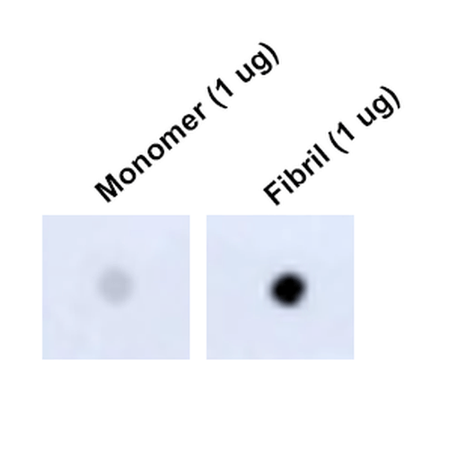 Dot Blot analysis using Mouse Anti-Tau Monoclonal Antibody, Clone 1D5 (57012). Tissue: Recombinant Protein. Species: Human. Primary Antibody: Mouse Anti-Tau Monoclonal Antibody (57012) at 1:1000 for 2 hours at RT with shaking. Secondary Antibody: Goat anti-mouse IgG:HRP at 1:5000 for 1 hour at RT with shaking.
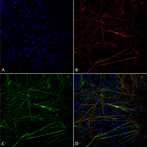 munocytochemistry/Immunofluorescence analysis using Mouse Anti-Tau Monoclonal Antibody, Clone 1D5 (57012). Tissue: iPSC-derived neurons. Species: Human. Fixation: 4% PFA. Primary Antibody: Mouse Anti-Tau Monoclonal Antibody (57012) at 1:100 for Overnight at 4°C. Counterstain: DAPI at 1:5000 for 5 minutes at RT in the dark. Magnification: 40X. Courtesy of: Francesco Paonessa.
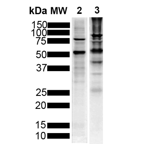 Western Blot analysis of Human Breast Cancer Cell line and Mouse Brain showing detection of Tau protein using Mouse Anti-Tau Monoclonal Antibody, Clone 1D5 (57012). Lane 1: MW Marker. Lane 2: Human T-47d (10ug). Lane 3: Mouse Brain (20ug). Block: 5% Skim Milk powder in TBST. Primary Antibody: Mouse Anti-Tau Monoclonal Antibody (57012) at 1:1000 for 2 hours at RT with shaking. Secondary Antibody: Goat anti-mouse IgG:HRP at 1:5000 for 1 hour at RT with shaking. Color Development: Chemiluminescent for HRP (Moss) for 5 min in RT.
 - -
- -
Antibody DetailsProduct DetailsReactive Species Human ⋅ Mouse ⋅ Rat Host Species Mouse Immunogen Full-length recombinant human Tau441 (2N4R) P301S mutant protein pre-formed fibrils. In humans, mutation P301S gives rise to early-onset dementia. Product Concentration 1.0 mg/ml Formulation PBS, pH 7.4, 0.09% sodium azide, 50% glycerol. State of Matter Liquid Product Preparation Purified by Protein G affinity chromatography Storage and Handling This product is stable for at least one (1) year at -20°C. Regulatory Status For in vitro investigational use only. Not intended for therapeutic or diagnostic procedures. Country of Origin USA Shipping Next Day 2-8°C Applications and Recommended Usage? Quality Tested by Leinco Immunoblotting: use at 1-5ug/mL. A band of ~45.8kDa is detected. Each investigator should determine their own optimal working dilution for specific applications. See directions on lot specific datasheets, as information may periodically change. DescriptionDescriptionSpecificity This antibody recognizes human, mouse, and rat Tau. Background Tau is a subunit protein of one of the major markers of Alzheimer disease (AD), the neurofibrillary
tangles. Tau protein has many unusual properties, such as its number of structural
conformations and biochemical modifications (phosphorylation, proteolysis, and glycosylation),
and its many interaction partners (mainly microtubules, but also other cytoskeletal proteins,
kinases, and phosphatases, motor proteins, chaperones, and membrane proteins). The
aggregation of Tau is toxic in cell and animal models, but can be reversed by suppressing
expression or by aggregation inhibitors. Function Promotes microtubule assembly and stability, and might be involved in the establishment and maintenance of neuronal polarity (PubMed:21985311). The C-terminus binds axonal microtubules while the N-terminus binds neural plasma membrane components, suggesting that tau functions as a linker protein between both (PubMed:21985311, PubMed:32961270). Axonal polarity is predetermined by TAU/MAPT localization (in the neuronal cell) in the domain of the cell body defined by the centrosome. The short isoforms allow plasticity of the cytoskeleton whereas the longer isoforms may preferentially play a role in its stabilization. {PubMed:21985311, PubMed:32961270}. NCBI Gene Bank ID UniProt.org Research Area Alzheimer's Disease References & CitationsTechnical Protocols ICC IF  Certificate of Analysis |
Formats Available
 Products are for research use only. Not for use in diagnostic or therapeutic procedures.