

Anti-Hsp70 [Polyclonal]

Data
 Immunocytochemistry/Immunofluorescence analysis using Rabbit Anti-Hsp70 Polyclonal Antibody (11108). Tissue: Heat Shocked Cervical cancer cell line (HeLa). Species: Human. Fixation: 2% Formaldehyde for 20 min at RT. Primary Antibody: Rabbit Anti-Hsp70 Polyclonal Antibody (11108) at 1:100 for 12 hours at 4°C. Secondary Antibody: FITC Goat Anti-Rabbit (green) at 1:200 for 2 hours at RT. Counterstain: DAPI (blue) nuclear stain at 1:40000 for 2 hours at RT. Localization: Cytoplasm. Magnification: 100x. (A) DAPI (blue) nuclear stain. (B) Anti-Hsp70 Antibody. (C) Composite. Heat Shocked at 42°C for 1h.
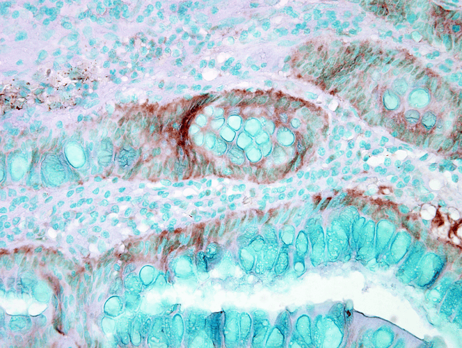 Immunohistochemistry analysis using Rabbit Anti-HSP70 Polyclonal Antibody (11108). Tissue: colon carcinoma. Species: Human. Fixation: Formalin. Primary Antibody: Rabbit Anti-HSP70 Polyclonal Antibody (11108) at 1:50000 for 12 hours at 4°C. Secondary Antibody: Biotin Goat Anti-Rabbit at 1:2000 for 1 hour at RT. Counterstain: Methyl Green at 200uL for 2 min at RT.
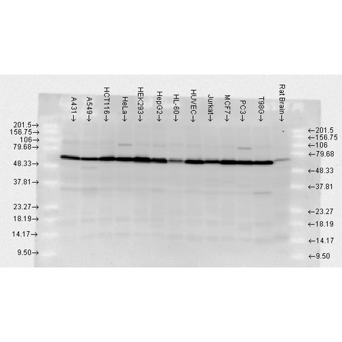 Western blot analysis of Human, Rat brain cell lysates showing detection of HSP70 protein using Rabbit Anti-HSP70 Polyclonal Antibody (11108). Load: 2 µg. Block: 1.5% BSA for 30 minutes at RT. Primary Antibody: Rabbit Anti-HSP70 Polyclonal Antibody (11108) at 1:10000 for 2 hours at RT. Secondary Antibody: Donkey Anti-Rabbit IgG: HRP for 1 hour at RT.
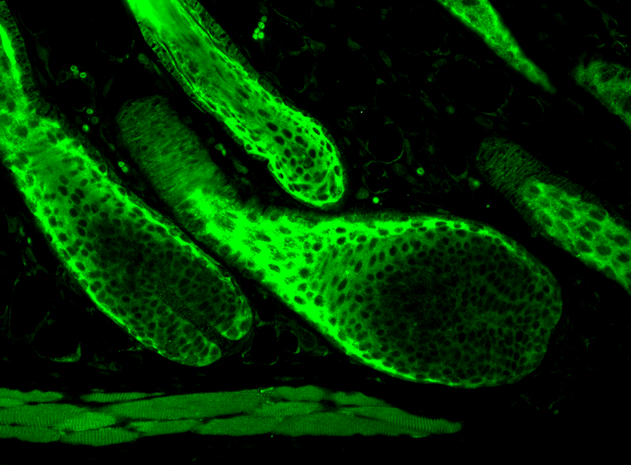 Immunohistochemistry analysis using Rabbit Anti-HSP70 Polyclonal Antibody (11108). Tissue: backskin. Species: Mouse. Fixation: Bouin’s Fixative Solution. Primary Antibody: Rabbit Anti-HSP70 Polyclonal Antibody (11108) at 1:100 for 1 hour at RT. Secondary Antibody: FITC Goat Anti-Rabbit (green) at 1:50 for 1 hour at RT. Localization: Cytoplasm.
 - -
- -
Antibody DetailsProduct DetailsReactive Species Beluga ⋅ Bovine ⋅ Canine ⋅ Coral ⋅ Fish ⋅ Guinea Pig ⋅ Hamster ⋅ Human ⋅ Monkey ⋅ Mouse ⋅ Ovine ⋅ Porcine ⋅ Rat Host Species Rabbit Immunogen Full-length Hsp70 protein. Product Concentration Lot Specific Formulation Whole antiserum State of Matter Liquid Product Preparation Whole antiserum Storage and Handling This antibody is stable for at least one (1) year at -20°C. Regulatory Status For in vitro investigational use only. Not
for use in therapeutic or diagnostic
procedures. Country of Origin USA Shipping Next Day 2-8°C Applications and Recommended Usage? Quality Tested by Leinco Immunoblotting: use at 1:10,000- 1:25,000 (ECL). A band of ~70 kDa is detected.
Immunoprecipitation: use at 1:100 dilution Positive control: HeLa cell lysate. Each investigator should determine their own optimal working dilution for specific applications. See directions on lot specific datasheets, as information may periodically change. DescriptionDescriptionSpecificity This antibody recognizes human,
mouse, rat, beluga, bovine, canine, fish,
guinea pig, hamster, monkey, porcine,
ovine, and coral Hsp70. Background Hsp70 binds ATP with high affinity and possesses a weak ATPase activity which can be stimulated by binding to unfolded proteins and synthetic peptides. Hsp70 recognizes and binds to nascent polypeptide chains as well as partially folded intermediates of proteins preventing their aggregation and misfolding. The binding of ATP triggers a critical conformational change leading to the release of the bound protein. The ability of Hsp70 to undergo cycles of binding and release from hydrophobic stretches of partially unfolded proteins is the basis for their role in a variety of intracellular functions such as protein synthesis, protein folding and oligomerization and protein transport. UniProt.org Research Area Heat Shock & Stress Proteins References & CitationsTechnical Protocols ICC IF    Certificate of Analysis |
