

Anti-Mouse PD-1 [Clone RMP1-30] BX027; Alexa Fluor™ 647-RX027


Anti-Mouse PD-1 [Clone RMP1-30] BX027; Alexa Fluor™ 647-RX027
Product No.: P415
- -
- -
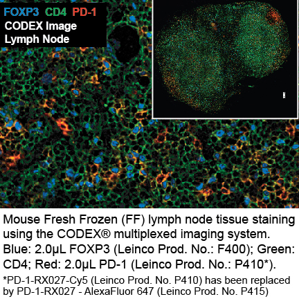
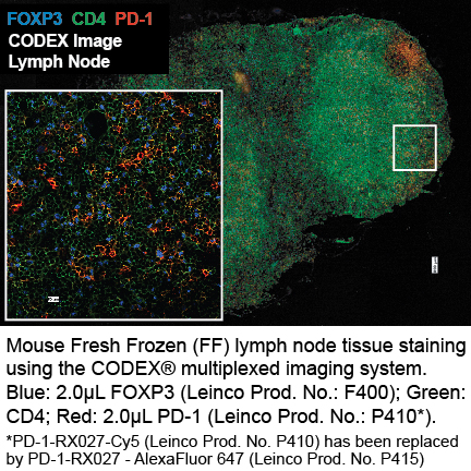
Clone RMP1-30 Target PD-1 Formats AvailableView All Product Type Monoclonal Antibody Alternate Names Programmed Death-1, CD279, PD 1 Isotype Rat IgG2a κ Applications IHC FF , PhenoCycler® |
Data
- -
- -
CODEX® DetailsBarcode BX027 Reporter AlexaFluor™ 647-RX027 Fluorescent Dye Alexa Fluor™ 647 is in the bright, far-red–fluorescent channel with an excitation max of 650 nm and an emission max of 671 nm. Tissue Screened MCA Sarcoma (Tumor) Tissue Preparation FF (Fresh Frozen) Antibody and Reporter DetailsReactivity Species Mouse Host Species Rat Immunogen Mouse PD-1 transfected BHK cells Formulation This PhenoCycler-Fusion (CODEX)® barcoded antibody is formulated in phosphate buffered saline (150 mM NaCl) PBS, EDTA pH 7.2 containing 0.09% sodium azide as a preservative. The CODEX® reporter is formulated in 1X Tris-EDTA (TE) pH 8.0 (10 mM Tris-HCl, 1 mM disodium EDTA, pH 8.0) Product Preparation Manufactured in an animal-free facility using only in vitro cell culture techniques, purified by affinity chromatography, and conjugated to a specific barcode under optimal conditions. State of Matter CODEX® barcoded antibody - Liquid; CODEX® reporter - Liquid Storage and Handling This PhenoCycler-Fusion (CODEX)® barcoded antibody is stable when stored at 2-8°C for up to 1 year (do not freeze). The CODEX® reporter is stable when frozen at -20°C for up to 1 year. Applications and Recommended Usage? Quality Tested by Leinco CODEX®; IHC FF - The suggested dilution for staining tissue in immunohistochemistry - Fresh Frozen (FF) is 1 μl of Anti-Mouse PD-1 (BX027) in a final volume of 200 μl of CODEX® staining buffer. RRID AB_2894157 Country of Origin USA Each investigator should determine their own optimal working dilution for specific applications. See directions on lot specific datasheets, as information may periodically change. DescriptionSpecificity Clone RMP1-30 recognizes an epitope on mouse PD-1. Antigen Distribution PD-1 is expressed on a subset of CD4-CD8- thymocytes, and on activated T and B cells. Background Antibody PD-1 is a 50-55 kD member of the B7 Ig superfamily. PD-1 is also a member of the extended CD28/CTLA-4 family of T cell regulators and is suspected to play a role in lymphocyte clonal selection and peripheral tolerance. The ligands of PD-1 are PD-L1 and PD-L2, and are also members of the B7 Ig superfamily. PD-1 and its ligands negatively regulate immune responses. PD-L1, or B7-Homolog 1, is a 40 kD type I transmembrane protein that has been reported to costimulate T cell growth and cytokine production. The interaction of PD-1 with its ligand PD-L1 is critical in the inhibition of T cell responses that include T cell proliferation and cytokine production. PD-L1 has increased expression in several cancers. Inhibition of the interaction between PD-1 and PD-L1 can serve as an immune checkpoint blockade by improving T-cell responses In vitro and mediating preclinical antitumor activity. Within the field of checkpoint inhibition, combination therapy using anti-PD1 in conjunction with anti-CTLA4 has significant therapeutic potential for tumor treatments. PD-L2 is a 25 kD type I transmembrane ligand of PD-1. Via PD-1, PD-L2 can serve as a coinhibitor of T cell functions. Regulation of T cell responses, including enhanced T cell proliferation and cytokine production, can result from mAbs that block the PD-L2 and PD-1 interaction. PhenoCycler-Fusion (CODEX)® reporters are made up of a fluorescent dye and a short oligonucleotide DNA barcode called a CODEX® Tag. Fluorescent reporters enable highly specific detection of corresponding barcodes, and the use of spectrally separated dyes allow for precise signal detection in up to three distinct fluorescence channels at one time. Antigen DetailsProtein Ligand/Receptor PD-L1 (B7-H1), PD-L2 Function Lymphocyte clonal selection, peripheral tolerance PubMed NCBI Gene Bank ID References & Citations1. Ardolino, M. et al. (2018) J Clin Invest. 128(10):4654-4668. PubMed
2. Schreiber, RD. et al. (2017) Cancer Immunol Res. 5(2):106-117. 3. Honjo, T. et al. (1992) EMBO J. 11:3887. Technical Protocols PhenoCycler® |
Formats Available
- -
- -
Prod No. | Description |
|---|---|
C3444 | |
C3452 | |
C3441 | |
C3446 | |
C3450 | |
C3442 | |
C3448 | |
P415 |
 Products are for research use only. Not for use in diagnostic or therapeutic procedures.