

Anti-Mouse PD-L1 [Clone 10F.9G2] — Purified in vivo GOLD™ Functional Grade
![Anti-Mouse PD-L1 [Clone 10F.9G2] — Purified in vivo GOLD™ Functional Grade, Parent vial — Leinco Prod. No. P363](https://www.leinco.com/wp-content/uploads/2025/09/P363-Anti-Mouse-PD-L1-Clone-10F.9G2-GOLD-Parent-Vial-500x500.jpg)

Anti-Mouse PD-L1 [Clone 10F.9G2] — Purified in vivo GOLD™ Functional Grade
Product No.: P363
Clone 10F.9G2 Target PD-L1 Formats AvailableView All Product Type Monoclonal Antibody Alternate Names B7-H1, CD274 Isotype Rat IgG2b κ Applications B , FC , IHC FF , in vivo , PhenoCycler® , WB |
Data
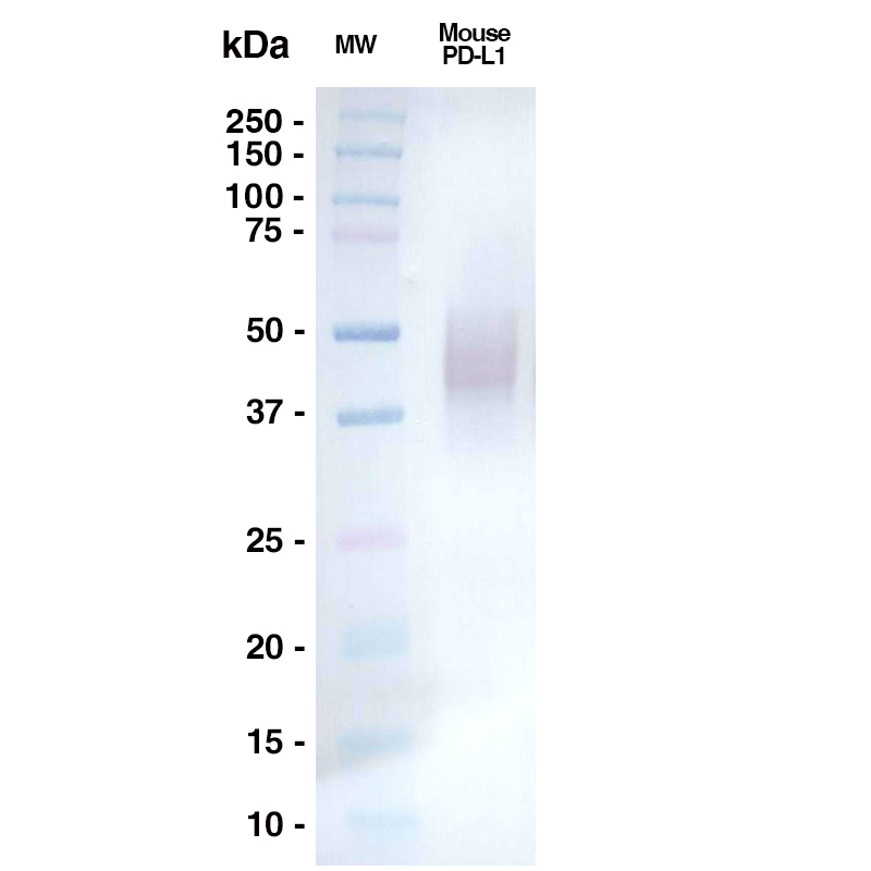 Western Blot
A 0.5ug sample of mouse PD-L1 (Leinco Prod. No.: P363) was loaded onto an SDS-PAGE gel under reducing conditions and probed with 10F.9G2 10ug/mL, Goat Anti-Rat IgG HRP (Goat Anti-Rat HRP (H+L) from Jackson Labs #112-035-062)
Antibody DetailsProduct DetailsReactive Species Mouse Host Species Rat Recommended Isotype Controls Recommended Dilution Buffer Immunogen UnKnown Product Concentration ≥ 5.0 mg/ml Endotoxin Level < 1.0 EU/mg as determined by the LAL method Purity ≥95% monomer by analytical SEC ⋅ >95% by SDS Page Formulation This monoclonal antibody is aseptically packaged and formulated in 0.01 M phosphate buffered saline (150 mM NaCl) PBS pH 7.2 - 7.4 with no carrier protein, potassium, calcium or preservatives added. Due to inherent biochemical properties of antibodies, certain products may be prone to precipitation over time. Precipitation may be removed by aseptic centrifugation and/or filtration. Product Preparation Functional grade preclinical antibodies are manufactured in an animal free facility using in vitro cell culture techniques and are purified by a multi-step process including the use of protein A or G to assure extremely low levels of endotoxins, leachable protein A or aggregates. Storage and Handling Functional grade preclinical antibodies may be stored sterile as received at 2-8°C for up to one month. For longer term storage, aseptically aliquot in working volumes without diluting and store at ≤ -70°C. Avoid Repeated Freeze Thaw Cycles. Country of Origin USA Shipping Next Day 2-8°C RRIDAB_2749826 Each investigator should determine their own optimal working dilution for specific applications. See directions on lot specific datasheets, as information may periodically change. DescriptionDescriptionSpecificity Clone 10F.9G2 recognizes an epitope on mouse PD-L1. Background PD-1 is a 50-55 kD member of the B7 Ig superfamily. PD-1 is also a member of the extended CD28/CTLA-4 family of T cell regulators and is suspected to play a role in lymphocyte clonal selection and peripheral tolerance. The ligands of PD-1 are PD-L1 and PD-L2, and are also members of the B7 Ig superfamily. PD-1 and its ligands negatively regulate immune responses. PD-L1, or B7-Homolog 1, is a 40 kD type I transmembrane protein that has been reported to costimulate T cell growth and cytokine production. The interaction of PD-1 with its ligand PD-L1 is critical in the inhibition of T cell responses that include T cell proliferation and cytokine production. PD-L1 has increased expression in several cancers. Inhibition of the interaction between PD-1 and PD-L1 can serve as an immune checkpoint blockade by improving T-cell responses In vitro and mediating preclinical antitumor activity. Within the field of checkpoint inhibition, combination therapy using anti-PD1 in conjunction with anti-CTLA4 has significant therapeutic potential for tumor treatments. PD-L2 is a 25 kD type I transmembrane ligand of PD-1. Via PD-1, PD-L2 can serve as a coinhibitor of T cell functions. Regulation of T cell responses, including enhanced T cell proliferation and cytokine production, can result from mAbs that block the PD-L2 and PD-1 interaction. Antigen Distribution PD-L1 is present on T cells, B cells, NK cells, dendritic cells, IFN-γ activated endothelial cells, and monocytes. Ligand/Receptor PD-1 (PDCD1) NCBI Gene Bank ID UniProt.org Research Area Cancer . Costimulatory Molecules . Immunology Leinco Antibody AdvisorPowered by AI: AI is experimental and still learning how to provide the best assistance. It may occasionally generate incorrect or incomplete responses. Please do not rely solely on its recommendations when making purchasing decisions or designing experiments. Clone 10F.9G2 is a rat monoclonal antibody targeting mouse PD-L1 (programmed death ligand 1) that has become a widely utilized tool in mouse immunology research. This antibody serves multiple critical functions in in vivo studies, particularly in understanding and manipulating immune checkpoint pathways. Blocking PD-L1 InteractionsThe primary in vivo application involves disrupting PD-L1 binding to its receptors, specifically PD-1 and B7-1 (CD80). By blocking these interactions, 10F.9G2 effectively removes inhibitory signals that normally suppress T cell activation and proliferation. This blocking mechanism operates in a dose-dependent manner and has been validated across numerous experimental systems. Cancer Immunotherapy ModelsIn oncology research, 10F.9G2 demonstrates significant antitumor activity. The antibody has been used to transiently arrest tumor growth in mouse models of melanoma by blocking the PD-L1/PD-1 interaction. This application makes it valuable for studying immune checkpoint blockade strategies and evaluating combination immunotherapy approaches in preclinical cancer models. Autoimmune Disease Studies10F.9G2 has proven particularly useful in autoimmune disease research, especially diabetes models. In NOD (non-obese diabetic) mice, administration of this antibody rapidly induces diabetes by unleashing diabetogenic effector T cells. In adoptive transfer experiments using CD8+ T cells from diabetic 8.3 NOD mice, treatment with 10F.9G2 resulted in 100% of recipient mice developing rapid-onset diabetes, compared to low incidence in control-treated animals. By day 11 post-transfer, approximately 40% of islets showed insulitis in 10F.9G2-treated mice versus less than 4% in controls. Immune Response ModulationBeyond specific disease models, 10F.9G2 serves as a general tool for detecting, modulating, or enhancing immune responses in mice. The antibody enables researchers to investigate how PD-L1-mediated signaling regulates T cell function, inflammation, and tolerance in various physiological and pathological contexts. This includes applications in studying dendritic cell function, T cell activation, and the balance between immune activation and suppression. Technical ConsiderationsFor in vivo applications, 10F.9G2 is available in specialized formulations designed for animal studies, with high purity (>95%) and low endotoxin levels (less than 1 EU/mg). These quality specifications are essential for minimizing non-specific inflammatory responses that could confound experimental results. The most commonly used antibodies or proteins with 10F.9G2 (anti-mouse PD-L1/B7-H1) in the literature are those that target related immune checkpoint pathways or immune cell markers, especially anti-PD-1 antibodies, other PD-L1 clones, and cell lineage markers. Commonly co-used antibodies and proteins include:
Researchers often use combinations of these antibodies to investigate immune checkpoint blockade, cell subset phenotyping, and detailed immune landscape characterization in murine models, particularly in studies of cancer immunotherapy and immune regulation. Clone 10F.9G2 is a widely used rat monoclonal antibody that specifically targets mouse PD-L1 (also known as CD274 or B7-H1), and it has been instrumental in elucidating the roles of PD-L1 in immune regulation, especially in mouse models. Key findings from scientific literature using citations of 10F.9G2 cluster around the following themes:
In summary, clone 10F.9G2 is a benchmark reagent in mouse immunology for probing and blocking PD-L1 function, and its dual blockade of PD-1 and B7-1 pathways has provided key insights into immune regulation and the basis for checkpoint inhibitor therapy in cancer. The dosing regimens for clone 10F.9G2, an anti-PD-L1 antibody, can vary across different mouse models. However, the standard dose range for this clone is typically between 100-250 μg per mouse. This is administered intraperitoneally, and the optimal dosing schedule is usually 2-3 times per week. The dosing regimens are influenced by the specific application, such as cancer immunotherapy studies or infection models, where the goal is to enhance T-cell responses by blocking the PD-1/PD-L1 interaction. One study mentions using a specific dose of 200 μg/mouse for PD-L1 blockade using 10F.9G2. Variations in dosing can depend on factors like the model's sensitivity to PD-L1 blockade, the presence of other immune modulators, and the desired immune response outcomes. In general, while the core dosing parameters remain consistent across models, specific dosing frequencies and amounts may be adjusted based on experimental objectives and the immunophenotypic characteristics of the models used, such as MC-38 and LLC1 tumor models. References & Citations1.) Ardolino, M. et al. (2018) J Clin Invest. 128(10):4654-4668. PubMed 2.) Schreiber, RD. et al. (2017) Cancer Immunol Res. 5(2):106-117. 3.) Gubin, M. et al. (2018) Cell. 175(4):1014–1030.e19 Journal Link Technical ProtocolsB  IHC FF  PhenoCycler®  Certificate of Analysis |
Related Products
Formats Available
Prod No. | Description |
|---|---|
P370 | |
P369 | |
P368 | |
P363 | |
P502 | |
P371 | |
P400 | |
P405 |
 Products are for research use only. Not for use in diagnostic or therapeutic procedures.