

Anti-Rat SOD2 (Mn)


Data
 Immunocytochemistry/Immunofluorescence analysis using Rabbit Anti-SOD (Mn) Polyclonal Antibody (13004). Tissue: Cervical cancer cell line (HeLa). Species: Human. Fixation: 2% Formaldehyde for 20 min at RT. Primary Antibody: Rabbit Anti-SOD (Mn) Polyclonal Antibody (13004) at 1:120 for 12 hours at 4°C. Secondary Antibody: R-PE Goat Anti-Rabbit (yellow) at 1:200 for 2 hours at RT. Counterstain: DAPI (blue) nuclear stain at 1:40000 for 2 hours at RT. Localization: Mitochondrion matrix. Magnification: 100x. (A) DAPI (blue) nuclear stain. (B) Anti-SOD (Mn) Antibody. (C) Composite.
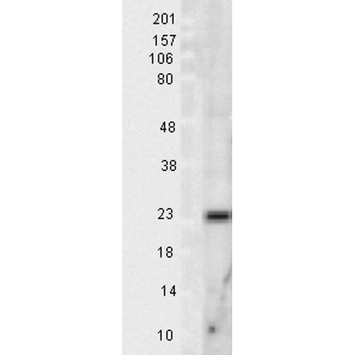 Western blot analysis of Rat Tissue lysates showing detection of SOD2 protein using Rabbit Anti-SOD2 Polyclonal Antibody (13004). Load: 15 µgprotein. Block: 1.5% BSA for 30 minutes at RT. Primary Antibody: Rabbit Anti-SOD2 Polyclonal Antibody (13004) at 1:1000 for 2 hours at RT. Secondary Antibody: Donkey Anti-Rabbit IgG: HRP for 1 hour at RT.
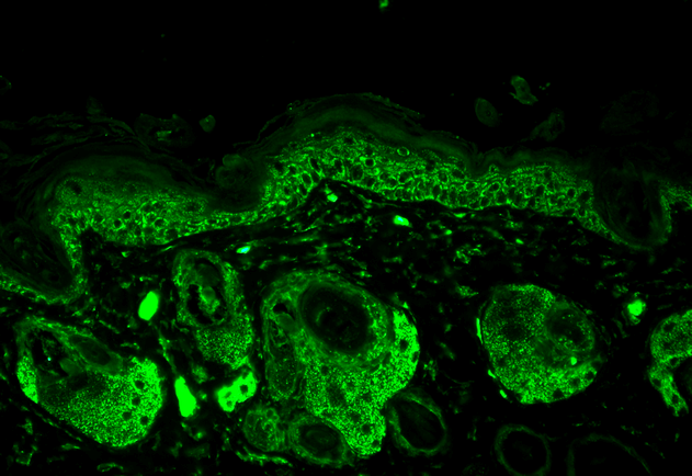 Immunohistochemistry analysis using Rabbit Anti-SOD2 Polyclonal Antibody (13004). Tissue: backskin. Species: Mouse. Fixation: Bouin’s Fixative Solution. Primary Antibody: Rabbit Anti-SOD2 Polyclonal Antibody (13004) at 1:100 for 1 hour at RT. Secondary Antibody: FITC Goat Anti-Rabbit (green) at 1:50 for 1 hour at RT. Localization: Mitochondrion matrix.
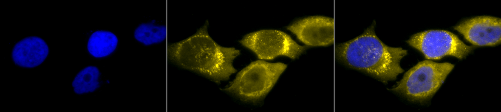
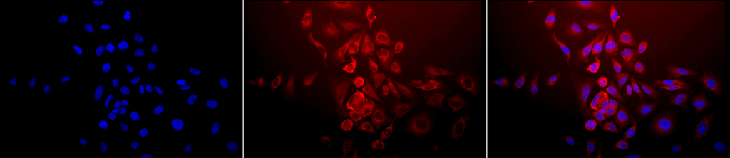 Immunocytochemistry/Immunofluorescence analysis using Rabbit Anti-SOD (Mn) Polyclonal Antibody (13004). Tissue: Cervical cancer cell line (HeLa). Species: Human. Fixation: 2% Formaldehyde for 20 min at RT. Primary Antibody: Rabbit Anti-SOD (Mn) Polyclonal Antibody (13004) at 1:120 for 12 hours at 4°C. Secondary Antibody: APC Goat Anti-Rabbit (red) at 1:200 for 2 hours at RT. Counterstain: DAPI (blue) nuclear stain at 1:40000 for 2 hours at RT. Localization: Mitochondrion matrix. Magnification: 20x. (A) DAPI (blue) nuclear stain. (B) Anti-SOD (Mn) Antibody. (C) Composite.
 - -
- -
Antibody DetailsProduct DetailsReactivity Species Bovine ⋅ Canine ⋅ Chicken ⋅ Gerbil ⋅ Guinea Pig ⋅ Hamster ⋅ Human ⋅ Monkey ⋅ Mouse ⋅ Porcine ⋅ Rabbit ⋅ Rat ⋅ Sheep ⋅ Xenopus Host Species Rabbit Immunogen Rat Mn SOD Product Concentration 1.0 mg/ml Formulation PBS, pH 7.0, 50% glycerol, and 0.09% sodium azide. State of Matter Liquid Product Preparation Purified by immunoaffinity chromatography Storage and Handling This antibody is stable for at least one (1) year at -20°C. Avoid multiple freeze-thaw cycles. Regulatory Status For in vitro investigational use only. Not intended for use in therapeutic or diagnostic procedures. Country of Origin USA Shipping Next Day 2-8°C Applications and Recommended Usage? Quality Tested by Leinco Immunoblotting: use at 0.5-1.0ug/mL
Immunohistochemistry: use at 1-10ug/mL These are recommended concentrations. Endusers should determine optimal concentrations for their applications. Each investigator should determine their own optimal working dilution for specific applications. See directions on lot specific datasheets, as information may periodically change. DescriptionSpecificity This antibody detects a 25kDa protein on SDS-PAGE immunoblots corresponding to the molecular mass of Mn SOD. This antibody recognizes human, monkey, mouse, rat, bovine, canine, porcine, hamster, gerbil, guinea pig, rabbit, sheep, chicken, and Xenopus Mn SOD. Background Superoxide dismutase (SOD) is an endogenously produced intracellular enzyme that catalyzes the dismutation of the superoxide radical O2- to oxygen and hydrogen peroxide which are then metabolized to H2O and O2 by catalase and glutathione peroxidase. SODs play an important role in antioxidant defense mechanisms. Three different SOD isoenzymes are found in mammalian cells: SOD1, SOD2, and SOD3. Antigen DetailsFunction Destroys superoxide anion radicals which are normally produced within the cells and which are toxic to biological systems. {PubMed:12791589}. NCBI Gene Bank ID UniProt.org Research Area Enzymes References & CitationsTechnical Protocols ICC IF    |
 Products are for research use only. Not for use in diagnostic or therapeutic procedures.