Anti-TSG101 Antibody (3352)
Anti-TSG101 Antibody (3352)
Product No.: 3352
- -
- -
Clone 4A10 Target TSG101 Formats AvailableView All Product Type Monoclonal Alternate Names ESCRT-I complex subunit TSG101 Isotype Mouse IgG1 Applications ELISA , EM , FACS , ICC , IF , IHC , IHC FFPE , IP , WB |
Data
![TSG101 antibody [4A10] detects TSG101 protein by western blot analysis. A. 30 ug NIH-3T3 whole cell lysate/extract B. 30 ug JC whole cell lysate/extract C. 30 ug BCL-1 whole cell lysate/extract 10% SDS-PAGE TSG101 antibody [4A10] (3352) dilution: 1:500 The HRP-conjugated anti-mouse IgG antibody was used to detect the primary antibody.](https://www.leinco.com/wp-content/uploads/2025/01/qed-bioscience-anti-tsg101-antibody-3352-1.jpg) TSG101 antibody [4A10] detects TSG101 protein by western blot analysis.
A. 30 ug NIH-3T3 whole cell lysate/extract
B. 30 ug JC whole cell lysate/extract
C. 30 ug BCL-1 whole cell lysate/extract
10% SDS-PAGE
TSG101 antibody [4A10] (3352) dilution: 1:500
The HRP-conjugated anti-mouse IgG antibody was used to detect the primary antibody.
TSG101 antibody [4A10] detects TSG101 protein by western blot analysis.
A. 30 ug NIH-3T3 whole cell lysate/extract
B. 30 ug JC whole cell lysate/extract
C. 30 ug BCL-1 whole cell lysate/extract
10% SDS-PAGE
TSG101 antibody [4A10] (3352) dilution: 1:500
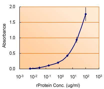
The HRP-conjugated anti-mouse IgG antibody was used to detect the primary antibody. ELISA detection of TSG101 using for capture at a concentration of 5 ug/mL and 3352 for detection at a concentration of 1.5 ug/mL.
![Various whole cell extracts (30 ug) were separated by 10% SDS-PAGE, and the membrane was blotted with TSG101 antibody [4A10] (3352) diluted at 1:500. The HRP-conjugated anti-mouse IgG antibody was used to detect the primary antibody.](https://www.leinco.com/wp-content/uploads/2025/01/qed-bioscience-anti-tsg101-antibody-3352-3.jpg) Various whole cell extracts (30 ug) were separated by 10% SDS-PAGE, and the membrane was blotted with TSG101 antibody [4A10] (3352) diluted at 1:500. The HRP-conjugated anti-mouse IgG antibody was used to detect the primary antibody.
Various whole cell extracts (30 ug) were separated by 10% SDS-PAGE, and the membrane was blotted with TSG101 antibody [4A10] (3352) diluted at 1:500. The HRP-conjugated anti-mouse IgG antibody was used to detect the primary antibody.![TSG101 antibody [4A10] detects TSG101 protein at cytoplasm by immunohistochemical analysis. Sample: Paraffin-embedded human breast carcinoma. TSG101 stained by TSG101 antibody [4A10] (3352) diluted at 1:100. Antigen Retrieval: Citrate buffer, pH 6.0, 15 min](https://www.leinco.com/wp-content/uploads/2025/01/qed-bioscience-anti-tsg101-antibody-3352-4.jpg) TSG101 antibody [4A10] detects TSG101 protein at cytoplasm by immunohistochemical analysis.
Sample: Paraffin-embedded human breast carcinoma.
TSG101 stained by TSG101 antibody [4A10] (3352) diluted at 1:100.
Antigen Retrieval: Citrate buffer, pH 6.0, 15 min
TSG101 antibody [4A10] detects TSG101 protein at cytoplasm by immunohistochemical analysis.
Sample: Paraffin-embedded human breast carcinoma.
TSG101 stained by TSG101 antibody [4A10] (3352) diluted at 1:100.
Antigen Retrieval: Citrate buffer, pH 6.0, 15 min![TSG101 antibody [4A10] detects TSG101 protein at cytoplasm by immunohistochemical analysis. Sample: Paraffin-embedded human ovarian cancer. TSG101 stained by TSG101 antibody [4A10] (3352) diluted at 1:100. Antigen Retrieval: Citrate buffer, pH 6.0, 15 min](https://www.leinco.com/wp-content/uploads/2025/01/qed-bioscience-anti-tsg101-antibody-3352-5.jpg) TSG101 antibody [4A10] detects TSG101 protein at cytoplasm by immunohistochemical analysis.
Sample: Paraffin-embedded human ovarian cancer.
TSG101 stained by TSG101 antibody [4A10] (3352) diluted at 1:100.
Antigen Retrieval: Citrate buffer, pH 6.0, 15 min
TSG101 antibody [4A10] detects TSG101 protein at cytoplasm by immunohistochemical analysis.
Sample: Paraffin-embedded human ovarian cancer.
TSG101 stained by TSG101 antibody [4A10] (3352) diluted at 1:100.
Antigen Retrieval: Citrate buffer, pH 6.0, 15 min![TSG101 antibody [4A10] (3352) detects TSG101 protein by flow cytometry analysis. Sample: THP-1 cell. Black: Unlabelled sample was used as a control. Red: TSG101 antibody [4A10] (3352) dilution: 1:25. Acquisition of 20,000 events were collected using a Dylight 488-conjugated secondary antibody for FACS analysis.](https://www.leinco.com/wp-content/uploads/2025/01/qed-bioscience-anti-tsg101-antibody-3352-6.jpg) TSG101 antibody [4A10] (3352) detects TSG101 protein by flow cytometry analysis.
Sample: THP-1 cell.
Black: Unlabelled sample was used as a control.
Red: TSG101 antibody [4A10] (3352) dilution: 1:25.
Acquisition of 20,000 events were collected using a Dylight 488-conjugated secondary antibody for FACS analysis.
TSG101 antibody [4A10] (3352) detects TSG101 protein by flow cytometry analysis.
Sample: THP-1 cell.
Black: Unlabelled sample was used as a control.
Red: TSG101 antibody [4A10] (3352) dilution: 1:25.
Acquisition of 20,000 events were collected using a Dylight 488-conjugated secondary antibody for FACS analysis.![Various whole cell extracts (30 ug) were separated by 10% SDS-PAGE, and the membrane was blotted with TSG101 antibody [4A10] (3352) diluted at 1:500. The HRP-conjugated anti-mouset IgG antibody was used to detect the primary antibody.](https://www.leinco.com/wp-content/uploads/2025/01/qed-bioscience-anti-tsg101-antibody-3352-7.jpg) Various whole cell extracts (30 ug) were separated by 10% SDS-PAGE, and the membrane was blotted with TSG101 antibody [4A10] (3352) diluted at 1:500. The HRP-conjugated anti-mouset IgG antibody was used to detect the primary antibody.
Various whole cell extracts (30 ug) were separated by 10% SDS-PAGE, and the membrane was blotted with TSG101 antibody [4A10] (3352) diluted at 1:500. The HRP-conjugated anti-mouset IgG antibody was used to detect the primary antibody.![Various tissue extracts (50 ug) were separated by 10% SDS-PAGE, and the membrane was blotted with TSG101 antibody [4A10] (3352) diluted at 1:500. The HRP-conjugated anti-mouse IgG antibody was used to detect the primary antibody.](https://www.leinco.com/wp-content/uploads/2025/01/qed-bioscience-anti-tsg101-antibody-3352-8.jpg) Various tissue extracts (50 ug) were separated by 10% SDS-PAGE, and the membrane was blotted with TSG101 antibody [4A10] (3352) diluted at 1:500. The HRP-conjugated anti-mouse IgG antibody was used to detect the primary antibody.
Various tissue extracts (50 ug) were separated by 10% SDS-PAGE, and the membrane was blotted with TSG101 antibody [4A10] (3352) diluted at 1:500. The HRP-conjugated anti-mouse IgG antibody was used to detect the primary antibody.![TSG101 antibody [4A10] detects TSG101 protein at cytoplasm by immunohistochemical analysis. Sample: Paraffin-embedded human lung cancer. TSG101 stained by TSG101 antibody [4A10] (3352) diluted at 1:50. Antigen Retrieval: Citrate buffer, pH 6.0, 15 min](https://www.leinco.com/wp-content/uploads/2025/01/qed-bioscience-anti-tsg101-antibody-3352-9.jpg) TSG101 antibody [4A10] detects TSG101 protein at cytoplasm by immunohistochemical analysis.
Sample: Paraffin-embedded human lung cancer.
TSG101 stained by TSG101 antibody [4A10] (3352) diluted at 1:50.
Antigen Retrieval: Citrate buffer, pH 6.0, 15 min
TSG101 antibody [4A10] detects TSG101 protein at cytoplasm by immunohistochemical analysis.
Sample: Paraffin-embedded human lung cancer.
TSG101 stained by TSG101 antibody [4A10] (3352) diluted at 1:50.
Antigen Retrieval: Citrate buffer, pH 6.0, 15 min - -
- -
Antibody DetailsProduct DetailsReactive Species Hamster ⋅ Human ⋅ Monkey ⋅ Mouse ⋅ Rat Host Species Mouse Immunogen Recombinant protein corresponding to aa 167-374 of TSG101 protein Product Concentration Lot Specific Formulation PBS, pH 7.2. State of Matter Liquid Product Preparation Purified by affinity chromatography Storage and Handling This product is stable for at least one (1) year if stored at -20°C. Store product in appropriate aliquots to avoid multiple freeze-thaw cycles. Regulatory Status Research Use Only Country of Origin USA Shipping Next Day 2-8°C Applications and Recommended Usage? Quality Tested by Leinco ELISA: use at 1-5ug/ml.
Immunoblotting: use at a 1:500-1:3,000 dilution. A band of 46kDa is detected. Detection of TSG101 protein in (A) NIH-3T3 cell lysate, (B) JC cell lysate, and (C) BCL-1 cell lysate with #3352 diluted 1:500. Endusers should determine optimal antibody concentrations for their applications. IHC: use at a dilution of 1:100 with antigen retrieval: citrate buffer, pH 6.0, 15 minutes. Detection of TSG101 protein in paraffin-embedded human breast carcinoma. Detection of TSG101 protein in paraffin-embedded human ovarian carcinoma. Each investigator should determine their own optimal working dilution for specific applications. See directions on lot specific datasheets, as information may periodically change. DescriptionDescriptionSpecificity This antibody recognizes human, mouse, rat, hamster, and monkey TSG101 protein. Background Tumor Susceptibility Gene 101 (TSG101) codes for a 46kDa protein whose inactivation in mouse
fibroblasts results in cell transformation and tumor formation in nude mice. Mutations in TSG101
have been linked to cervical, breast, prostate and gastrointestinal cancers. In addition, TSG101
is one of the cellular proteins involved in the budding process of HIV-1, and plays an important
role in the cellular vacuolar protein sorting (Vps) pathway. Its main function being recognizing
ubiquitinated cargo, TSG101 also proved to be essential for the budding process of HIV-1 virions. Function Component of the ESCRT-I complex, a regulator of vesicular trafficking process. Binds to ubiquitinated cargo proteins and is required for the sorting of endocytic ubiquitinated cargos into multivesicular bodies (MVBs). Mediates the association between the ESCRT-0 and ESCRT-I complex. Required for completion of cytokinesis; the function requires CEP55. May be involved in cell growth and differentiation. Acts as a negative growth regulator. Involved in the budding of many viruses through an interaction with viral proteins that contain a late-budding motif P-[ST]-A-P. This interaction is essential for viral particle budding of numerous retroviruses. Required for the exosomal release of SDCBP, CD63 and syndecan (PubMed:22660413). It may also play a role in the extracellular release of microvesicles that differ from the exosomes (PubMed:22315426). {PubMed:11916981, PubMed:17556548, PubMed:17853893, PubMed:21070952, PubMed:21757351, PubMed:22315426, PubMed:22660413}. NCBI Gene Bank ID UniProt.org Research Area Cancer Research References & CitationsTechnical ProtocolsCertificate of Analysis |


![Various whole cell extracts (30 ug) were separated by 10% SDS-PAGE, and the membrane was blotted with TSG101 antibody [4A10] (3352) diluted at 1:500. The HRP-conjugated anti-mouse IgG antibody was used to detect the primary antibody.](https://www.leinco.com/wp-content/uploads/2025/01/qed-bioscience-anti-tsg101-antibody-3352-10.jpg)
![Various whole cell extracts (30 ug) were separated by 10% SDS-PAGE, and the membrane was blotted with TSG101 antibody [4A10] (3352) diluted at 1:500. The HRP-conjugated anti-mouse IgG antibody was used to detect the primary antibody, and the signal was developed with Trident ECL plus-Enhanced.](https://www.leinco.com/wp-content/uploads/2025/01/qed-bioscience-anti-tsg101-antibody-3352-11.jpg)
