

Anti-Dityrosine (DT) [Clone 10A6]


Anti-Dityrosine (DT) [Clone 10A6]
Product No.: 56622
- -
- -
Clone 10A6 Target Dityrosine Formats AvailableView All Product Type Monoclonal Alternate Names Dityrosine, Dityrosine (DT), DT, DY Isotype Mouse IgG1 Applications ELISA , FACS , ICC , IF , WB , FCM |
Data
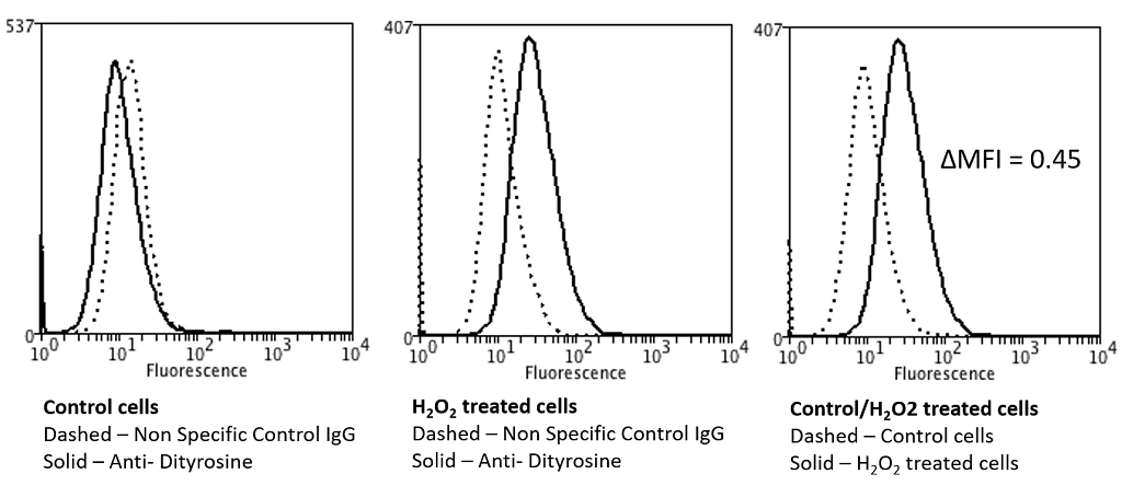 Flow Cytometry analysis using Mouse Anti-Dityrosine Monoclonal Antibody, Clone 10A6 . Tissue: Neuroblastoma cells (SH-SY5Y). Species: Human. Fixation: 90% Methanol. Primary Antibody: Mouse Anti-Dityrosine Monoclonal Antibody at 1:50 for 30 min on ice. Secondary Antibody: Goat Anti-Mouse: PE at 1:100 for 20 min at RT. Isotype Control: Non Specific IgG. Cells were subject to oxidative stress by treating with 250 µM H2O2 for 24 hours.
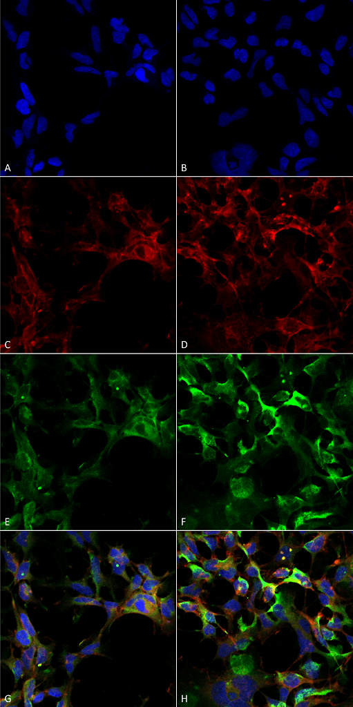 Immunocytochemistry/Immunofluorescence analysis using Mouse Anti-Dityrosine Monoclonal Antibody, Clone 10A6 . Tissue: Embryonic kidney epithelial cell line (HEK293). Species: Human. Fixation: 5% Formaldehyde for 5 min. Primary Antibody: Mouse Anti-Dityrosine Monoclonal Antibody at 1:50 for 30-60 min at RT. Secondary Antibody: Goat Anti-Mouse Alexa Fluor 488 at 1:1500 for 30-60 min at RT. Counterstain: Phalloidin Alexa Fluor 633 F-Actin stain; DAPI (blue) nuclear stain at 1:250, 1:50000 for 30-60 min at RT. Localization: Cytoplasmic. Magnification: 20X (2X Zoom). (A,C,E,G) – Untreated. (B,D,F,H) – Cells cultured overnight with 50 µM H2O2. (A,B) DAPI (blue) nuclear stain. (C,D) Phalloidin Alexa Fluor 633 F-Actin stain. (E,F) Dityrosine Antibody. (G,H) Composite. Courtesy of: Dr. Robert Burke, University of Victoria.
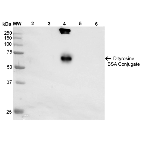 Western Blot analysis of Dityrosine-BSA Conjugate showing detection of 67 kDa Dityrosine protein using Mouse Anti-Dityrosine Monoclonal Antibody, Clone 10A6 . Lane 1: Molecular Weight Ladder (MW). Lane 2: BSA. Lane 3: 3,5-Dibromotyrosine-BSA. Lane 4: Dityrosine-BSA. Lane 5: Bromotyrosine-BSA. Lane 6: 7-ketocholesterol-BSA. Load: 1 µg. Block: 5% Skim Milk in TBST. Primary Antibody: Mouse Anti-Dityrosine Monoclonal Antibody at 1:1000 for 2 hours at RT. Secondary Antibody: Goat Anti-Mouse IgG: HRP at 1:2000 for 60 min at RT. Color Development: ECL solution for 5 min in RT. Predicted/Observed Size: 67 kDa.
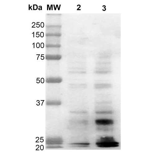 Western Blot analysis of Human Cervical cancer cell line (HeLa) lysate showing detection of Dityrosine protein using Mouse Anti-Dityrosine Monoclonal Antibody, Clone 10A6 . Lane 1: Molecular Weight Ladder (MW). Lane 2: HeLa cell lysate. Lane 3: H2O2 treated HeLa cell lysate. Load: 12 µg. Block: 5% Skim Milk in TBST. Primary Antibody: Mouse Anti-Dityrosine Monoclonal Antibody at 1:1000 for 2 hours at RT. Secondary Antibody: Goat Anti-Mouse IgG: HRP at 1:2000 for 60 min at RT. Color Development: ECL solution for 5 min in RT.
 - -
- -
Antibody DetailsProduct DetailsReactive Species Species Independent Host Species Mouse Immunogen Synthetic Dityrosine conjugated to Keyhole Limpet Hemocyanin (KLH). Product Concentration 1 mg/mL Formulation PBS pH 7.4, 50% glycerol, 0.09% Sodium azide State of Matter Liquid Product Preparation Protein G Purified Storage and Handling This antibody is stable for at least one (1) year at -20°C. Avoid multiple freeze-thaw cycles. Regulatory Status Research Use Only Country of Origin USA Shipping Next Day 2-8°C Applications and Recommended Usage? Quality Tested by Leinco WB (1:1000); ICC/IF (1:50); FACS (1:50); FCM (1:50); ELISA (1:1000); optimal dilutions for assays should be determined by the user. Each investigator should determine their own optimal working dilution for specific applications. See directions on lot specific datasheets, as information may periodically change. DescriptionDescriptionSpecificity Specific for dityrosine modified proteins. Does not cross-react with 3,5-dibromotyrosine or bromotyrosine modified proteins. Background ROS such as hydrogen peroxide (H2O2), superoxide, and hydroxyl radicals can react with both the backbone and the side chains of proteins, leading to backbone cleavage and side-chain modifications, respectively. Peroxidases, UV radiation, and hydroxyl radicals catalyze the formation of tyrosyl radicals which then react to form cross-links between proteins (1). This produces dityrosine, a metabolically stable biomarker of protein oxidation (2). Research Area Cancer . Cell Signaling . Oxidation . Oxidative Stress . Post-translational Modifications . Protein Oxidation References & CitationsTechnical Protocols FACS ICC IF  FCM Certificate of Analysis |
Formats Available
 Products are for research use only. Not for use in diagnostic or therapeutic procedures.