

Anti-HSF1 [Clone 10H8]


Anti-HSF1 [Clone 10H8]
Product No.: 12518
- -
- -
Clone 10H8 Target HSF1 Formats AvailableView All Product Type Monoclonal Alternate Names HSF 1, Heat shock transcription factor 1, HSTF 1 Isotype Rat IgG1 Applications ELISA , ICC , IF , IHC , IP , WB , GS |
Data
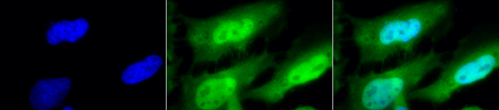 Immunocytochemistry/Immunofluorescence analysis using Rat Anti-HSF1 Monoclonal Antibody, Clone 10H8 (12518). Tissue: Heat Shocked cervical cancer cells (HeLa). Species: Human. Fixation: 2% Formaldehyde for 20 min at RT. Primary Antibody: Rat Anti-HSF1 Monoclonal Antibody (12518) at 1:100 for 12 hours at 4°C. Secondary Antibody: FITC Goat Anti-Rat (green) at 1:200 for 2 hours at RT. Counterstain: DAPI (blue) nuclear stain at 1:40000 for 2 hours at RT. Localization: Diffuse nuclear and cytoplasmic staining. Magnification: 100x. (A) DAPI (blue) nuclear stain. (B) Anti-HSF1 Antibody. (C) Composite. Heat Shocked at 42°C for 1h.
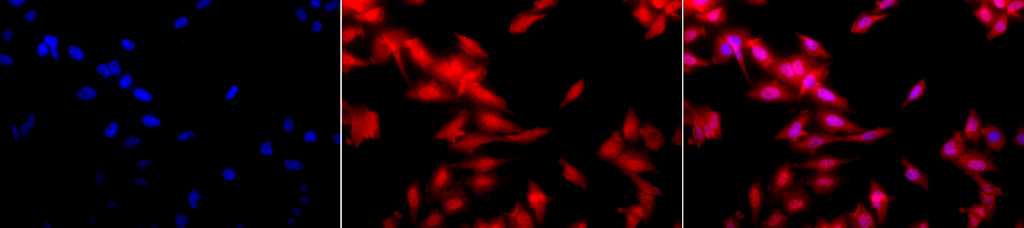 Immunocytochemistry/Immunofluorescence analysis using Rat Anti-HSF1 Monoclonal Antibody, Clone 10H8 (12518). Tissue: Heat Shocked cervical cancer cells (HeLa). Species: Human. Fixation: 2% Formaldehyde for 20 min at RT. Primary Antibody: Rat Anti-HSF1 Monoclonal Antibody (12518) at 1:100 for 12 hours at 4°C. Secondary Antibody: APC Goat Anti-Rat (red) at 1:200 for 2 hours at RT. Counterstain: DAPI (blue) nuclear stain at 1:40000 for 2 hours at RT. Localization: Diffuse nuclear and cytoplasmic staining. Magnification: 20x. (A) DAPI (blue) nuclear stain. (B) Anti-HSF1 Antibody. (C) Composite. Heat Shocked at 42°C for 1h.
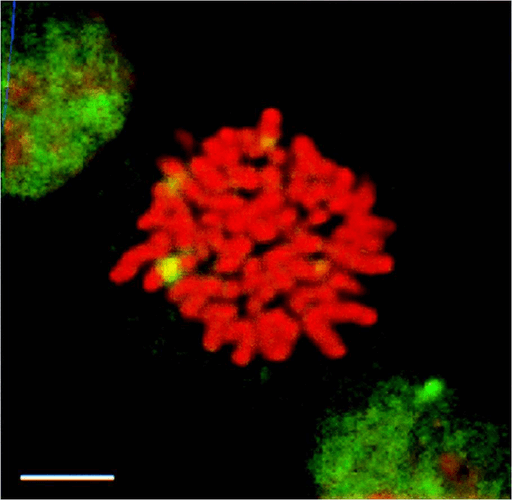 Immunocytochemistry/Immunofluorescence analysis using Rat Anti-HSF1 Monoclonal Antibody, Clone 10H8 (12518). Tissue: Heat Shocked mitotic HeLa cells. Species: Human. Primary Antibody: Rat Anti-HSF1 Monoclonal Antibody (12518) at 1:1000. HSF1 stained green. Courtesy of: Morimoto Lab, Northwestern University, USA.
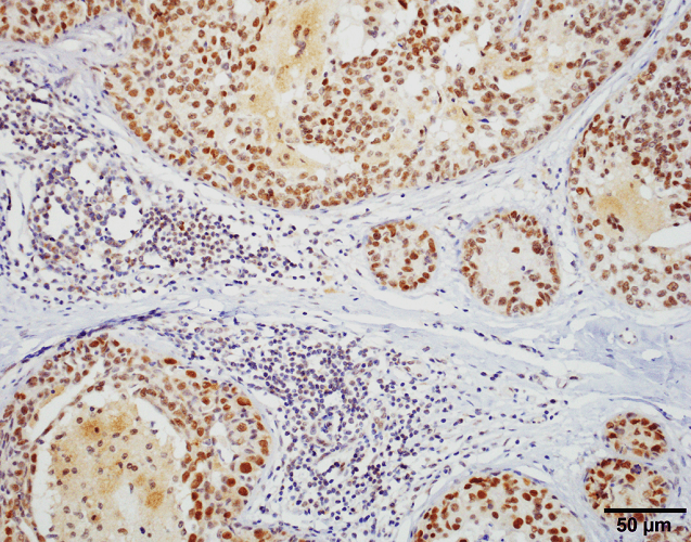 Immunohistochemistry analysis using Rat Anti-HSF1 Monoclonal Antibody, Clone 10H8 (12518). Tissue: Breast carcinoma. Species: Human. Fixation: 10% Formalin Solution for 20 hours at RT. Primary Antibody: Rat Anti-HSF1 Monoclonal Antibody (12518) at 1:1000 for 40 min. Secondary Antibody: Dako labeled Polymer HRP Anti-rat IgG, DAB Chromogen (brown) (Dako Envision+ System) for 30 min at RT. Counterstain: Mayer’s Hematoxylin (purple/blue) nuclear stain for 1 minute at RT. Localization: Nuclear. Magnification: 100X. Courtesy of: Dr. Sandro Santagata, Harvard Medical School.
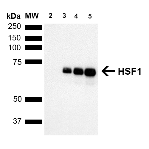 Western Blot analysis of Human Breast adenocarcinoma cell line (MCF7) showing detection of ~65 kDa HSF1 protein using Rat Anti-HSF1 Monoclonal Antibody, Clone 10H8 (12518). Lane 1: MW ladder. Lane 2: HSF1 null lysate prepared from mouse embryonic fibroblasts. Lane 3: MCF7 lysate (5 µg). Lane 4: MCF7 lysate (10 µg). Lane 5: MCF7 lysate (20 µg). Block: 1.5% BSA for 30 minutes at RT. Primary Antibody: Rat Anti-HSF1 Monoclonal Antibody (12518) at 1:1000 for 2 hours at RT. Secondary Antibody: Goat Anti-Rat IgG: HRP for 1 hour at RT. Predicted/Observed Size: ~65 kDa. Courtesy of: Dr. Sandro Santagata, Harvard Medical School.
 - -
- -
Antibody DetailsProduct DetailsReactivity Species Bovine ⋅ Guinea Pig ⋅ Hamster ⋅ Human ⋅ Monkey ⋅ Mouse ⋅ Rabbit ⋅ Rat Host Species Rat Immunogen Recombinant mouse HSF-1 Product Concentration Lot Specific Formulation PBS, pH 7.4. State of Matter Liquid Product Preparation Purified by Protein G affinity chromatography Storage and Handling This antibody is stable for at least one (1) year at -20°C. Avoid repeated freeze-thaw cycles. Regulatory Status For in vitro investigational use only. Not for use in therapeutic or diagnostic procedures. Country of Origin USA Shipping Next Day 2-8°C Applications and Recommended Usage? Quality Tested by Leinco Immunoblotting: use at 1-2ug/mL. A band of ~85 and/or ~95 kDa is detected.
Immunocytochemistry: use at 10ug/mL. Immunoprecipitation: use at 12.5ug/mL These are recommended concentrations. User should determine optimal concentrations for their application. Positive control: HeLa cell lysate Each investigator should determine their own optimal working dilution for specific applications. See directions on lot specific datasheets, as information may periodically change. DescriptionSpecificity This antibody recognizes an ~85 kDa
protein (inactive form) in unstressed cell
lysates and an ~95 kDa protein (active form)
in heat-shocked cell lysates of human,
mouse, rat, rabbit, bovine, guinea pig,
hamster, and monkey. Background Heat shock factor 1 (HSF1) is a heat shock transcription factor that activates the transcription of genes encoding products required for protein folding, processing, targeting, degradation, and function. Up- regulation of expression of heat shock proteins in response to stress occurs at the level of transcription through a heat shock element and a HSF transcription factor. Amino acid sequences for most HSFs are highly conserved. A DNA binding domain is at the N-terminus, hydrophobic repeats (essential to the formation of active trimers) are adjacent to this binding domain, and another short hydrophobic repeat (necessary for suppression of trimerization) occurs toward the C-terminus. In higher eukaryotes, HSF1 is distributed diffusely in the cell cytoplasm and nucleus in unstressed cells. On exposure to heat shock or other stresses, HSF1 localizes to discrete nuclear granules; on recovery from stress, it returns to a diffuse nuclear- cytoplasmic distribution. Antigen DetailsFunction Functions as a stress-inducible and DNA-binding transcription factor that plays a central role in the transcriptional activation of the heat shock response (HSR), leading to the expression of a large class of molecular chaperones heat shock proteins (HSPs) that protect cells from cellular insults' damage (PubMed:1871105, PubMed:11447121, PubMed:1986252, PubMed:7760831, PubMed:7623826, PubMed:8946918, PubMed:8940068, PubMed:9341107, PubMed:9121459, PubMed:9727490, PubMed:9499401, PubMed:9535852, PubMed:12659875, PubMed:12917326, PubMed:15016915, PubMed:25963659, PubMed:26754925, PubMed:18451878). In unstressed cells, is present in a HSP90-containing multichaperone complex that maintains it in a non-DNA-binding inactivated monomeric form (PubMed:9727490, PubMed:11583998, PubMed:16278218). Upon exposure to heat and other stress stimuli, undergoes homotrimerization and activates HSP gene transcription through binding to site-specific heat shock elements (HSEs) present in the promoter regions of HSP genes (PubMed:1871105, PubMed:1986252, PubMed:8455624, PubMed:7935471, PubMed:7623826, PubMed:8940068, PubMed:9727490, PubMed:9499401, PubMed:10359787, PubMed:11583998, PubMed:12659875, PubMed:16278218, PubMed:25963659, PubMed:26754925). Upon heat shock stress, forms a chromatin-associated complex with TTC5/STRAP and p300/EP300 to stimulate HSR transcription, therefore increasing cell survival (PubMed:18451878). Activation is reversible, and during the attenuation and rPubMed:10359787, PubMed:11447121, PubMed:11583998, PubMed:12659875, PubMed:12917326, PubMed:14707147, PubMed:15016915, PubMed:16278218, PubMed:17897941, PubMed:18451878, PubMed:1871105, PubMed:18794143, PubMed:1986252, PubMed:25963659, PubMed:26359349, PubMed:26727489, PubMed:26754925, PubMed:7623826, PubMed:7760831, PubMed:7935471, PubMed:8455624, PubMed:8940068, PubMed:8946918, PubMed:9121459, PubMed:9341107, PubMed:9499401, PubMed:9535852, PubMed:9727490}.; (Microbial infection) Plays a role in latent human immunodeficiency virus (HIV-1) transcriptional reactivation. Binds to the HIV-1 long terminal repeat promoter (LTR) to reactivate viral transcription by recruiting cellular transcriptional elongation factors, such as CDK9, CCNT1 and EP300. {PubMed:27189267}. NCBI Gene Bank ID UniProt.org Research Area Heat Shock & Stress Proteins References & CitationsCotto, J et al. 1997 J Cell Science 110: 2925-2934. Technical Protocols ICC IF    GS |
Formats Available
 Products are for research use only. Not for use in diagnostic or therapeutic procedures.