Anti-Hsp90α [Clone D7α]
Anti-Hsp90α [Clone D7α]
Product No.: 11102
- -
- -
Clone D7alpha Target Hsp90α Formats AvailableView All Product Type Monoclonal Isotype Mouse IgG1 Applications ELISA , IHC , IP , WB , AM |
Data
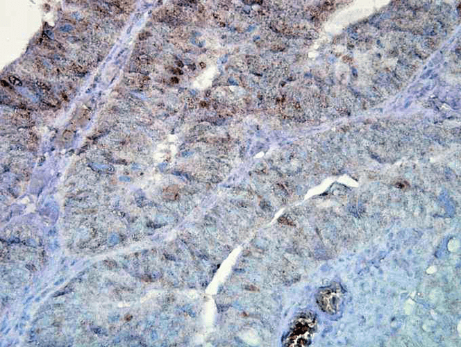
 Immunohistochemistry analysis using Mouse Anti-Hsp90 Monoclonal Antibody, Clone D7alpha (11102). Tissue: colon carcinoma. Species: Human. Fixation: Formalin. Primary Antibody: Mouse Anti-Hsp90 Monoclonal Antibody (11102) at 1:100000 for 12 hours at 4°C. Secondary Antibody: Biotin Goat Anti-Mouse at 1:2000 for 1 hour at RT. Counterstain: Mayer Hematoxylin (purple/blue) nuclear stain at 200 µl for 2 minutes at RT. Magnification: 40x. This image was produced using an amplifying IHC wash buffer. The antibody has therefore been diluted more than is recommended for other applications.
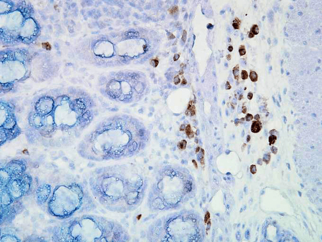
 Immunohistochemistry analysis using Mouse Anti-Hsp90 Monoclonal Antibody, Clone D7alpha (11102). Tissue: inflamed colon. Species: Mouse. Fixation: Formalin. Primary Antibody: Mouse Anti-Hsp90 Monoclonal Antibody (11102) at 1:100000 for 12 hours at 4°C. Secondary Antibody: Biotin Goat Anti-Mouse at 1:2000 for 1 hour at RT. Counterstain: Mayer Hematoxylin (purple/blue) nuclear stain at 200 µl for 2 minutes at RT. Localization: Inflammatory cells. Magnification: 40x. Inflammatory cells. This image was produced using an amplifying IHC wash buffer. The antibody has therefore been diluted more than is recommended for other applications.
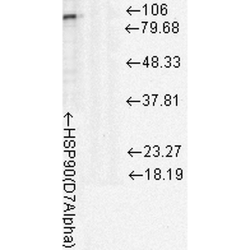
 Western Blot analysis of Rat cell lysates showing detection of Hsp90 protein using Mouse Anti-Hsp90 Monoclonal Antibody, Clone D7Alpha (11102). Load: 15 µg. Block: 1.5% BSA for 30 minutes at RT. Primary Antibody: Mouse Anti-Hsp90 Monoclonal Antibody (11102) at 1:1000 for 2 hours at RT. Secondary Antibody: Sheep Anti-Mouse IgG: HRP for 1 hour at RT.
 - -
- -
Antibody DetailsProduct DetailsReactivity Species Bovine ⋅ Chicken ⋅ Human ⋅ Mouse ⋅ Porcine ⋅ Rabbit ⋅ Rat Host Species Mouse Immunogen Full-length Hsp90 purified from chicken brain Product Concentration Lot Specific Formulation PBS, pH 7.4. State of Matter Liquid Product Preparation Purified by Protein G affinity chromatography Storage and Handling This antibody is stable for at least one (1) year at -20°C. This antibody recognizes human, mouse, rat, rabbit, bovine, porcine, and chicken Hsp90alpha (90 kDa). Regulatory Status For in vitro investigational use only. Not for
use in therapeutic or diagnostic procedures. Country of Origin USA Shipping Next Day 2-8°C Applications and Recommended Usage? Quality Tested by Leinco Immunoblotting: use at 1-5ug/mL. A band of ~90 kDa is detected.
Immunoprecipitation: 5ug on 20ul Protein A-Sepharose + 100ul sample. These are recommended concentrations. User should determine optimal concentrations for their application. Positive control: Heat-shocked HeLa cell lysate. Each investigator should determine their own optimal working dilution for specific applications. See directions on lot specific datasheets, as information may periodically change. DescriptionSpecificity This antibody recognizes human, mouse, rat, rabbit, bovine, porcine, and chicken Hsp90alpha (90 kDa). Background Hsp90 and the 94 kDa glucose-regulated protein, Grp94, are major molecular chapeones of the cytosol and endoplasmic reticulum. In mammalian cells, there are at least two Hsp90 isoforms, Hsp90alpha and Hsp90beta, which are encoded by separate genes. All known members of the Hsp90 family are highly conserved, especially in the N-terminal and C-terminal regions. In the absence of stress, Hsp90 is an essential component of cellular processes such as hormone signaling and cell cycle control. Several regulatory proteins such as steroid receptors, cell cycle kinases and p53 have been identified as substrates of Hsp90. Antigen DetailsFunction Molecular chaperone that promotes the maturation, structural maintenance and proper regulation of specific target proteins involved for instance in cell cycle control and signal transduction. Undergoes a functional cycle that is linked to its ATPase activity which is essential for its chaperone activity. This cycle probably induces conformational changes in the client proteins, thereby causing their activation. Interacts dynamically with various co-chaperones that modulate its substrate rUniProtKB:P07900}. NCBI Gene Bank ID UniProt.org Research Area Heat Shock & Stress Proteins References & CitationsSchuh S et al. 1985 J Biol Chem 260: 14292-14296 Sullivan WP et al. 1985 Biochemistry 24: 4214-4222. Technical Protocols |


