

Anti-Mouse/Human CD273 (PD-L2) [Clone 3.2.B8] — Purified in vivo GOLD™ Functional Grade


Anti-Mouse/Human CD273 (PD-L2) [Clone 3.2.B8] — Purified in vivo GOLD™ Functional Grade
Product No.: P379
Clone 3.2.B8 Target PD-L2 Formats AvailableView All Product Type Monoclonal Antibody Alternate Names B7-DC, CD273, PDL2, B7DC, Clone 3.2 Isotype Mouse IgG1 Applications B , FC , in vivo , WB |
Data
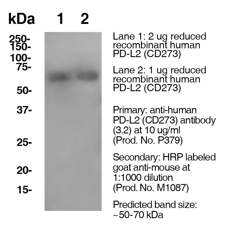
Antibody DetailsProduct DetailsReactive Species Human ⋅ Mouse Host Species Mouse Recommended Isotype Controls Recommended Isotype Controls Recommended Dilution Buffer Immunogen Made in PD-L2 knockout mouse fusion partner X63-Ag8.653 myeloma cells Product Concentration ≥ 5.0 mg/ml Endotoxin Level < 1.0 EU/mg as determined by the LAL method Purity ≥95% monomer by analytical SEC ⋅ >95% by SDS Page Formulation This monoclonal antibody is aseptically packaged and formulated in 0.01 M phosphate buffered saline (150 mM NaCl) PBS pH 7.2 - 7.4 with no carrier protein, potassium, calcium or preservatives added. Due to inherent biochemical properties of antibodies, certain products may be prone to precipitation over time. Precipitation may be removed by aseptic centrifugation and/or filtration. Product Preparation Functional grade preclinical antibodies are manufactured in an animal free facility using in vitro cell culture techniques and are purified by a multi-step process including the use of protein A or G to assure extremely low levels of endotoxins, leachable protein A or aggregates. Storage and Handling Functional grade preclinical antibodies may be stored sterile as received at 2-8°C for up to one month. For longer term storage, aseptically aliquot in working volumes without diluting and store at ≤ -70°C. Avoid Repeated Freeze Thaw Cycles. Country of Origin USA Shipping Next Day 2-8°C RRIDAB_2737559 Applications and Recommended Usage? Quality Tested by Leinco Western Blot: For Western blotting, the suggested use of this reagent is 10 µg per ml (See Data Results) Each investigator should determine their own optimal working dilution for specific applications. See directions on lot specific datasheets, as information may periodically change. DescriptionDescriptionSpecificity Clone 3.2.B8 recognizes an epitope on mouse/human PD-L2 and has been shown to bind to both mouse and human PD-L2 equally well Background PD-1 is a 50-55 kD member of the B7 Ig superfamily. PD-1 is also a member of the extended CD28/CTLA-4 family of T cell regulators and is suspected to play a role in lymphocyte clonal selection and peripheral tolerance. The ligands of PD-1 are PD-L1 and PD-L2, and are also members of the B7 Ig superfamily. PD-1 and its ligands negatively regulate immune responses. PD-L1, or B7-Homolog 1, is a 40 kD type I transmembrane protein that has been reported to costimulate T cell growth and cytokine production. The interaction of PD-1 with its ligand PD-L1 is critical in the inhibition of T cell responses that include T cell proliferation and cytokine production. PD-L1 has increased expression in several cancers. Inhibition of the interaction between PD-1 and PD-L1 can serve as an immune checkpoint blockade by improving T-cell responses In vitro and mediating preclinical antitumor activity. Within the field of checkpoint inhibition, combination therapy using anti-PD1 in conjunction with anti-CTLA4 has significant therapeutic potential for tumor treatments. PD-L2 is a 25 kD type I transmembrane ligand of PD-1. Via PD-1, PD-L2 can serve as a co-inhibitor of T cell functions. Regulation of T cell responses, including enhanced T cell proliferation and cytokine production, can result from mAbs that block the PD-L2 and PD-1 interaction. Antigen Distribution PD-L2 is expressed on dendritic cells, liver, few transformed cell lines, and a subset of macrophages. Ligand/Receptor PD-1, uncharacterized receptor Function Binds to PD-1 and alternative receptor; NCBI Gene Bank ID UniProt.org Research Area Costimulatory Molecules . Immunology Leinco Antibody AdvisorPowered by AI: AI is experimental and still learning how to provide the best assistance. It may occasionally generate incorrect or incomplete responses. Please do not rely solely on its recommendations when making purchasing decisions or designing experiments. Clone 3.2.B8 is a monoclonal antibody targeting PD-L2 (CD273), used in in vivo mouse studies to modulate immune responses by blocking the interaction between PD-L2 and PD-1. This blockade plays a role in enhancing T cell activation, proliferation, and cytokine production, potentially leading to increased antitumor immunity. Key points on in vivo usage:
Additional details:
In summary, clone 3.2.B8 is employed in vivo mainly for immune checkpoint blockade studies in mice, with its primary purpose being to modulate T cell responses by interfering with PD-L2/PD-1 signaling. The 3.2.B8 antibody commonly refers to a clone specific for mouse CD273 (PD-L2), used in immunological research, especially for studying immune checkpoints and T cell regulation. In published studies, 3.2.B8 is often used alongside other antibodies or proteins to provide comprehensive immunophenotyping or functional analysis. Commonly co-used antibodies/proteins with 3.2.B8 (anti-PD-L2) include:
These combinations allow researchers to map out immune cell interactions, checkpoint blockade responses, and specific immune cell populations within in vivo or in vitro systems. In summary, 3.2.B8 (anti-PD-L2) is most frequently used with antibodies targeting PD-1, PD-L1, and standard immunophenotyping markers (CD3, CD4, CD8, CD11c, MHC-II), depending on the precise research application. The exact panel will vary by experiment, but these are the most recurrently reported combinations in immunological literature. Scientific literature referencing clone 3.2.B8 primarily identifies this monoclonal antibody as a highly specific tool for recognizing PD-L2 (CD273) on both mouse and human cells. The key findings from these citations include:
Applications in Research:
Experimental Utility:
No studies from the retrieved results link clone 3.2.B8 directly to infectious disease or TCR clonotype analysis, as discussed in related HIV T cell literature. Instead, its major scientific impact is enabling robust investigations into the function and therapeutic targeting of the PD-1/PD-L2 axis in immune modulation, including autoimmunity and oncology. If you need details on specific experimental findings or therapeutic outcomes using clone 3.2.B8, additional targeted literature searches may be necessary. The dosing regimens of clone 3.2.B8 across different mouse models are not directly specified in the available search results, and no published protocols are found that use clone 3.2.B8 by name. Based on typical practices for in vivo antibody dosing in mice, dosing regimens can vary significantly by mouse strain, research objective, and biological target. For antibodies in mouse models:
Key factors affecting dosing choice:
If you are planning to use clone 3.2.B8 specifically:
No direct information for clone 3.2.B8 appears in the cited literature, so adopt dosing regimens from antibodies with similar mechanisms and targets, modifying for mouse model idiosyncrasies. Always validate tolerability and efficacy in preliminary experiments. References & Citations1.) Akbari O, Stock P, Singh AK, Lombardi V, Lee WL, Freeman GJ, Sharpe AH, Umetsu
DT, Dekruyff RH. PD-L1 and PD-L2 modulate airway inflammation and iNKT-cell-
dependent airway hyperreactivity in opposing directions. Mucosal Immunol. 2010; 3:81-
91. PMCID: PMC2845714 2.) Xiao Y, Yu S, Zhu B, Bedoret D, Bu X, Francisco LM, Hua P, Duke-Cohan JS, Umetsu DT, Sharpe AH, DeKruyff RH*, Freeman GJ* (* indicates co-senior authors). RGMb is a novel binding partner for PD-L2 and its engagement with PD-L2 promotes respiratory tolerance. J Exp Med. 2014; 211:943-59. PMCID: PMC4010901. Technical ProtocolsB    Certificate of Analysis |
Related Products
Prod No. | Description |
|---|---|
S211 | |
R1364 | |
I-536 | |
M1188 | |
C247 | |
F1175 | |
S571 |
Formats Available
 Products are for research use only. Not for use in diagnostic or therapeutic procedures.