

Anti-Malondialdehyde [Clone 11E3]


Anti-Malondialdehyde [Clone 11E3]
Product No.: 56616
- -
- -
Clone 11E3 Target Malondialdehyde Formats AvailableView All Product Type Monoclonal Alternate Names Malondialdehyde, MDA, Malondialdehyde (MDA), Malonic aldehyde Propanedial, 1,3-Propanedial, Malonaldehyde Isotype Mouse IgG1 Applications ELISA , ICC , IF , IHC , WB |
Data
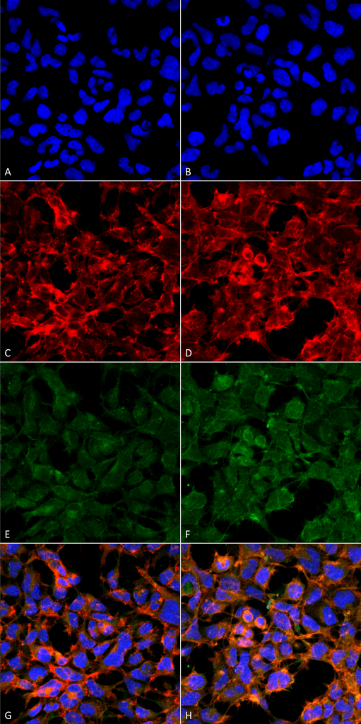 Immunocytochemistry/Immunofluorescence analysis using Mouse Anti-Malondialdehyde Monoclonal Antibody, Clone 11E3 . Tissue: Embryonic kidney epithelial cell line (HEK293). Species: Human. Fixation: 5% Formaldehyde for 5 min. Primary Antibody: Mouse Anti-Malondialdehyde Monoclonal Antibody at 1:50 for 30-60 min at RT. Secondary Antibody: Goat Anti-Mouse Alexa Fluor 488 at 1:1500 for 30-60 min at RT. Counterstain: Phalloidin Alexa Fluor 633 F-Actin stain; DAPI (blue) nuclear stain at 1:250, 1:50000 for 30-60 min at RT. Magnification: 20X (2X Zoom). (A,C,E,G) – Untreated. (B,D,F,H) – Cells cultured overnight with 50 µM H2O2. (A,B) DAPI (blue) nuclear stain. (C,D) Phalloidin Alexa Fluor 633 F-Actin stain. (E,F) Malondialdehyde Antibody. (G,H) Composite. Courtesy of: Dr. Robert Burke, University of Victoria.
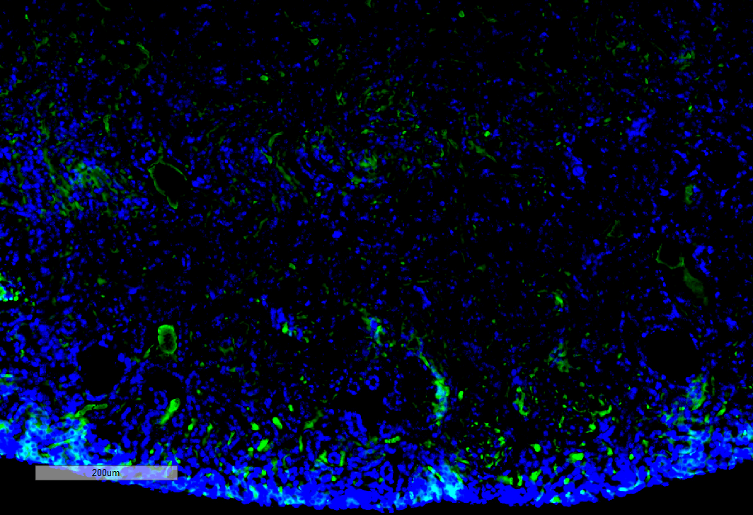 Immunohistochemistry analysis using Mouse Anti-Malondialdehyde Monoclonal Antibody, Clone 11E3 . Tissue: Kidney. Species: Mouse. Primary Antibody: Mouse Anti-Malondialdehyde Monoclonal Antibody at 1:100 for Overnight at 4C, then 30 min at 37C. Secondary Antibody: Goat Anti-Mouse IgG (H+L): FITC for 45 min at 37C. Counterstain: DAPI for 3 min at RT. Magnification: 10X.
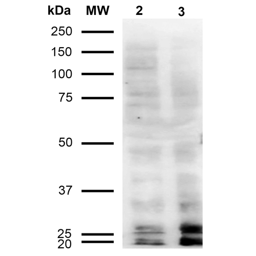 Western Blot analysis of Human Cervical cancer cell line (HeLa) lysate showing detection of Malondialdehyde protein using Mouse Anti-Malondialdehyde Monoclonal Antibody, Clone 11E3 . Lane 1: Molecular Weight Ladder (MW). Lane 2: HeLa cell lysate. Lane 3: H2O2 treated HeLa cell lysate. Load: 12 µg. Block: 5% Skim Milk in TBST. Primary Antibody: Mouse Anti-Malondialdehyde Monoclonal Antibody at 1:1000 for 2 hours at RT. Secondary Antibody: Goat Anti-Mouse IgG: HRP at 1:2000 for 60 min at RT. Color Development: ECL solution for 5 min in RT.
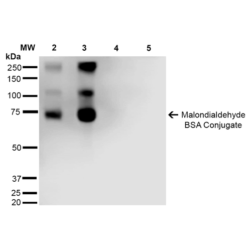 Western Blot analysis of Malondialdehyde-BSA Conjugate showing detection of 67 kDa Malondialdehyde protein using Mouse Anti-Malondialdehyde Monoclonal Antibody, Clone 11E3 . Lane 1: Molecular Weight Ladder (MW). Lane 2: Malondialdehyde-BSA (0.5 µg). Lane 3: Malondialdehyde-BSA (2.0 µg). Lane 4: BSA (0.5 µg). Lane 5: BSA (2.0 µg) . Block: 5% Skim Milk in TBST. Primary Antibody: Mouse Anti-Malondialdehyde Monoclonal Antibody at 1:1000 for 2 hours at RT. Secondary Antibody: Goat Anti-Mouse IgG: HRP at 1:2000 for 60 min at RT. Color Development: ECL solution for 5 min in RT. Predicted/Observed Size: 67 kDa.
 - -
- -
Antibody DetailsProduct DetailsReactivity Species Species Independent Host Species Mouse Immunogen Synthetic Malondialdehyde modified Keyhole Limpet Hemocyanin (KLH). Product Concentration 1 mg/mL Formulation PBS pH 7.4, 50% glycerol, 0.09% Sodium azide State of Matter Liquid Product Preparation Protein G Purified Storage and Handling This antibody is stable for at least one (1) year at -20°C. Avoid multiple freeze-thaw cycles. Regulatory Status Research Use Only Country of Origin USA Shipping Next Day 2-8°C Applications and Recommended Usage? Quality Tested by Leinco WB (1:1000); ICC/IF (1:50); ELISA (1:1000); IHC (1:100); Optimal dilutions for assays should be determined by the user. Each investigator should determine their own optimal working dilution for specific applications. See directions on lot specific datasheets, as information may periodically change. DescriptionSpecificity Specific for Malondialdehyde conjugated proteins. Does not detect free Malondialdehyde. Does not cross-react with Acrolein, Crotonaldehyde, Hexanoyl Lysine, 4-Hydroxy-2-hexenal, 4-Hydroxy nonenal, or Methylglyoxal modified proteins. Background Malondialdehyde (MDA) is the biomarker in greatest diagnostic use, due to its molecular stability. This three-carbon, low-molecular weight aldehyde has a strong affinity for amino acids, which results in adduct formation to both free amino acids and proteins. Increased MDA levels have been found at correlating levels in breast cancer, and lung cancer patients. Other diseased states with elevated MDA levels include diabetes and Alzheimer’s disease. Multiple laboratory techniques exist for quantification of MDA levels, including the thiobarbituric acid reactive substances (TBARS) assay. In addition to use as a biomarker, MDA has been shown to have mutagenic effects on tissues themselves as adduct formation can result in DNA cross-linking. Antigen DetailsResearch Area Cancer . Neuroscience . Alzheimer's Disease . Lipid peroxidation . Neurodegeneration . Oxidative Stress References & CitationsTechnical Protocols ICC IF   |
Formats Available
 Products are for research use only. Not for use in diagnostic or therapeutic procedures.