

Anti-Acrolein [Clone 10A10]


Anti-Acrolein [Clone 10A10]
Product No.: 56606
- -
- -
Clone 10A10 Target Acrolein Formats AvailableView All Product Type Monoclonal Alternate Names Acrolein modified protein, Acrolein conjugated protein, 2-Propen-1-one, 2-propenal, Acraldehyde, Acrolein, Acrylic aldehyde, Protein-bound Acrolein Isotype Mouse IgG1 Applications ELISA , FACS , ICC , IF , WB , FCM |
Data
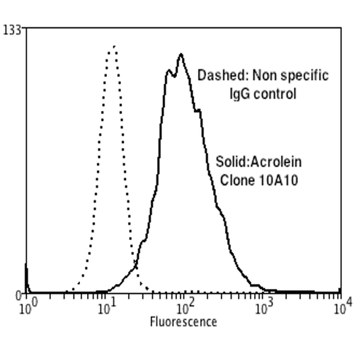 Flow Cytometry analysis using Mouse Anti-Acrolein Monoclonal Antibody, Clone 10A10 . Tissue: Neuroblastoma cells (SH-SY5Y). Species: Human. Fixation: 90% Methanol. Primary Antibody: Mouse Anti-Acrolein Monoclonal Antibody at 1:50 for 30 min on ice. Secondary Antibody: Goat Anti-Mouse: PE at 1:100 for 20 min at RT. Isotype Control: Non Specific IgG. Cells were subject to oxidative stress by treating with 250 µM H2O2 for 24 hours.
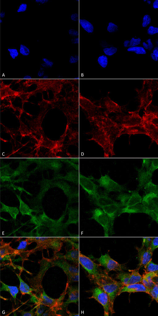 Immunocytochemistry/Immunofluorescence analysis using Mouse Anti-Acrolein Monoclonal Antibody, Clone 10A10 . Tissue: Embryonic kidney epithelial cell line (HEK293). Species: Human. Fixation: 5% Formaldehyde for 5 min. Primary Antibody: Mouse Anti-Acrolein Monoclonal Antibody at 1:50 for 30-60 min at RT. Secondary Antibody: Goat Anti-Mouse Alexa Fluor 488 at 1:1500 for 30-60 min at RT. Counterstain: Phalloidin Alexa Fluor 633 F-Actin stain; DAPI (blue) nuclear stain at 1:250, 1:50000 for 30-60 min at RT. Magnification: 20X (2X Zoom). (A,C,E,G) – Untreated. (B,D,F,H) – Cells cultured overnight with 50 µM H2O2. (A,B) DAPI (blue) nuclear stain. (C,D) Phalloidin Alexa Fluor 633 F-Actin stain. (E,F) Acrolein Antibody. (G,H) Composite. Courtesy of: Dr. Robert Burke, University of Victoria.
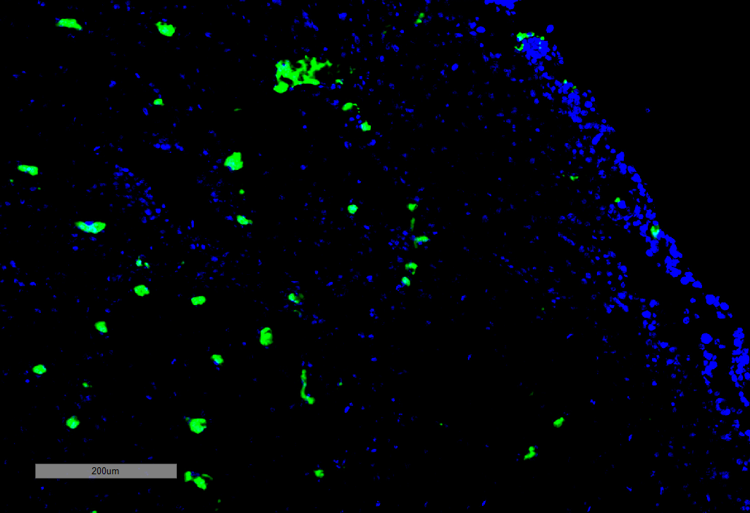 Immunohistochemistry analysis using Mouse Anti-Acrolein Monoclonal Antibody, Clone 10A10 . Tissue: Adrenal Carcinoma. Species: Human. Primary Antibody: Mouse Anti-Acrolein Monoclonal Antibody at 1:100 for Overnight at 4C, then 30 min at 37C. Secondary Antibody: Goat Anti-Mouse IgG (H+L): FITC for 45 min at 37C. Counterstain: DAPI for 3 min at RT. Magnification: 5X.
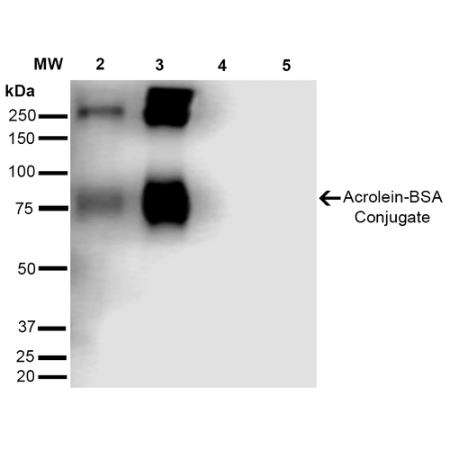 Western Blot analysis of Acrolein-BSA Conjugate showing detection of 67 kDa Acrolein protein using Mouse Anti-Acrolein Monoclonal Antibody, Clone 10A10 . Lane 1: Molecular Weight Ladder (MW). Lane 2: AcroleinBSA (0.5 µg). Lane 3: AcroleinBSA (2.0 µg). Lane 4: BSA (0.5 µg). Lane 5: BSA (2.0 µg). Block: 5% Skim Milk in TBST. Primary Antibody: Mouse Anti-Acrolein Monoclonal Antibody at 1:1000 for 2 hours at RT. Secondary Antibody: Goat Anti-Mouse IgG: HRP at 1:2000 for 60 min at RT. Color Development: ECL solution for 5 min in RT. Predicted/Observed Size: 67 kDa.
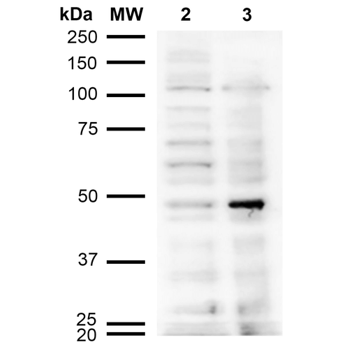 Western Blot analysis of Human Cervical cancer cell line (HeLa) lysate showing detection of Acrolein protein using Mouse Anti-Acrolein Monoclonal Antibody, Clone 10A10 . Lane 1: Molecular Weight Ladder (MW). Lane 2: HeLa cell lysate. Lane 3: H2O2 treated HeLa cell lysate. Load: 12 µg. Block: 5% Skim Milk in TBST. Primary Antibody: Mouse Anti-Acrolein Monoclonal Antibody at 1:1000 for 2 hours at RT. Secondary Antibody: Goat Anti-Mouse IgG: HRP at 1:2000 for 60 min at RT. Color Development: ECL solution for 5 min in RT.
 - -
- -
Antibody DetailsProduct DetailsReactivity Species Species Independent Host Species Mouse Immunogen Synthetic Acrolein modified Keyhole Limpet Hemocyanin (KLH). Product Concentration 1 mg/mL Formulation PBS pH 7.4, 50% glycerol, 0.09% Sodium azide State of Matter Liquid Product Preparation Protein G Purified Storage and Handling This antibody is stable for at least one (1) year at -20°C. Avoid multiple freeze-thaw cycles. Regulatory Status Research Use Only Country of Origin USA Shipping Next Day 2-8°C Applications and Recommended Usage? Quality Tested by Leinco WB (1:1000); ICC/IF (1:50); FACS (1:50); FCM (1:50); ELISA (1:1000); IHC (1:100); Optimal dilutions for assays should be determined by the user. Each investigator should determine their own optimal working dilution for specific applications. See directions on lot specific datasheets, as information may periodically change. DescriptionSpecificity Specific for Acrolein modified proteins. Does not detect free acrolein. Does not cross-react with Crotonaldehyde, Hexanoyl Lysine, 4-Hydroxy-2-hexenal, 4-Hydroxy nonenal, Malondialdehyde, or Methylglyoxal modified proteins. Background Lipid peroxidation occurs when oxidizing agents attack carbon-carbon double bonds found in unsaturated lipids. In addition to membrane degradation, oxidation end-products have been found to damage cell viability through their mutagenic and toxic properties. These downstream functional consequences facilitate the development of disease and premature aging. Acrolein is an electrophilic conjugated aldehyde that is a terminal product of lipid peroxidation. Acrolein is highly mutagenic and reacts with nucleophilic functional groups in DNA and proteins such as cysteine, histidine, and lysine residues (1).
Antigen DetailsResearch Area Cancer . Neuroscience . Alzheimer's Disease . Lipid peroxidation . Neurodegeneration . Oxidative Stress References & CitationsTechnical Protocols FACS ICC IF  FCM |
Formats Available
 Products are for research use only. Not for use in diagnostic or therapeutic procedures.