

Anti-Mouse CD86 [Clone GL1] — Purified in vivo GOLD™ Functional Grade


Anti-Mouse CD86 [Clone GL1] — Purified in vivo GOLD™ Functional Grade
Product No.: C2158
Clone GL1 Target B7-2 Formats AvailableView All Product Type Monoclonal Antibody Alternate Names B7-2, B70, Ly-58, CD-86 Isotype Rat IgG2a κ Applications B , ELISA , IHC FF , in vivo , IP , WB |
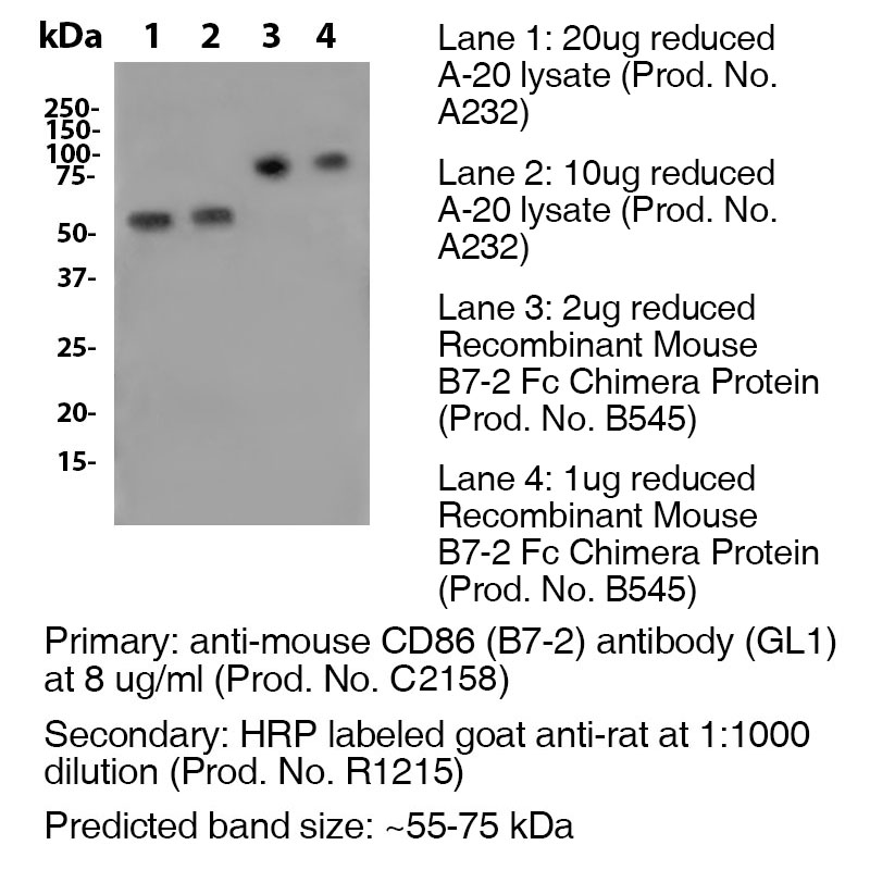
Data
Antibody DetailsProduct DetailsReactive Species Mouse Host Species Rat Recommended Isotype Controls Recommended Isotype Controls Recommended Dilution Buffer Immunogen LPS-activated CBA/Ca mouse splenic B cells Product Concentration ≥ 5.0 mg/ml Endotoxin Level < 1.0 EU/mg as determined by the LAL method Purity ≥95% monomer by analytical SEC ⋅ >95% by SDS Page Formulation This monoclonal antibody is aseptically packaged and formulated in 0.01 M phosphate buffered saline (150 mM NaCl) PBS pH 7.2 - 7.4 with no carrier protein, potassium, calcium or preservatives added. Due to inherent biochemical properties of antibodies, certain products may be prone to precipitation over time. Precipitation may be removed by aseptic centrifugation and/or filtration. Product Preparation Functional grade preclinical antibodies are manufactured in an animal free facility using in vitro cell culture techniques and are purified by a multi-step process including the use of protein A or G to assure extremely low levels of endotoxins, leachable protein A or aggregates. Storage and Handling Functional grade preclinical antibodies may be stored sterile as received at 2-8°C for up to one month. For longer term storage, aseptically aliquot in working volumes without diluting and store at ≤ -70°C. Avoid Repeated Freeze Thaw Cycles. Country of Origin USA Shipping Next Day 2-8°C RRIDAB_2749827 Each investigator should determine their own optimal working dilution for specific applications. See directions on lot specific datasheets, as information may periodically change. DescriptionDescriptionSpecificity Clone GL-1 recognizes an epitope on mouse CD86. Background CD86 is an 80kD Ig superfamily member that is involved in immunoglobulin class-switching and activation of NK cell-mediated cytotoxicity. CD80 is closely related to, and works in tandem with CD86 to prime T- cells. CD86 is expressed earlier in the immune response than CD80. The ligation of CD28 on T cells with CD80 and CD86 on APCs co-stimulates T cells resulting in enhanced cell activation, proliferation, and cytokine production. CD86 can also bind to CTLA-4 to deliver an inhibitory signal to T cells. Antigen Distribution CD86 is expressed on activated B and T cells, macrophages, dendritic cells, and astrocytes. Ligand/Receptor CD28, CD152 (CTLA-4) Function T cell costimulation, Ig class-switching, NK cell cytotoxicity PubMed NCBI Gene Bank ID UniProt.org Research Area Cell Biology . Costimulatory Molecules . Immunology . Neuroscience . Neuroscience Cell Markers Leinco Antibody AdvisorPowered by AI: AI is experimental and still learning how to provide the best assistance. It may occasionally generate incorrect or incomplete responses. Please do not rely solely on its recommendations when making purchasing decisions or designing experiments. Clone GL1 is a monoclonal antibody targeting mouse CD86 (B7-2) that is widely used in in vivo mouse studies to block costimulatory signaling necessary for T cell activation and immune responses. In vivo, administration of GL1 has been shown to inhibit the priming of cytotoxic T lymphocytes, especially when used in combination with antibodies against CD80 (B7-1). Supporting details:
Additional information:
In summary, clone GL1 is primarily used in in vivo mouse studies to block CD86-dependent costimulatory signaling, leading to inhibition of T cell-mediated immune responses and facilitating mechanistic studies of immune regulation. Commonly used antibodies or proteins studied with GL1 depend on the context, as "GL1" may refer to the glucose transporter 1 protein (GLUT1) or the glucagon-like peptide-1 receptor (GLP-1R). In the literature, several antibodies and proteins are frequently used alongside these targets for experimental and therapeutic research. If GL1 refers to GLUT1 (Glucose Transporter 1):
If GL1 refers to GLP-1R (Glucagon-Like Peptide-1 Receptor):
Additional Relevant Proteins/Antibodies:
Summary Table: Common Antibodies/Proteins Used with GLUT1 or GLP-1R
The choice of co-used antibodies or proteins should always be informed by the specific target and research objective. If a more specific or alternative meaning for "GL1" was intended, please clarify for a more tailored answer. Key findings from scientific literature citing clone GL1 (frequently in the context of GLP-1 production clones or analogues) focus on advances in recombinant production, bioactivity optimization, and molecular engineering for therapeutic applications, particularly in diabetes and obesity. Essential Findings:
Broader Context:
If you are seeking references to a different meaning for "clone GL1" (e.g., plant biology or unrelated molecular clones), please specify the field for more targeted information. The research cited here is focused on the recombinant production of glucagon-like peptide-1 (GLP-1) analogues. Dosing regimens for clone GL1, which targets mouse CD86 (B7-2), are not comprehensively detailed in the provided search results—most sources mention GL1's target specificity but lack concrete dosing protocols in mouse models. Regimens for similar monoclonal antibodies in mouse models, such as checkpoint inhibitors, often use doses in the range of 100–250 µg per mouse, administered intraperitoneally 2–3 times per week for cancer immunotherapy or infection models. Essential context and details:
Limitations:
Recommendation:
If you require detailed GL1 regimens for a specific disease model or mouse strain, consulting primary protocols from recent publications using GL1 or reaching out to antibody vendors/manufacturers is advised, since they sometimes provide technical sheets or usage references. References & Citations1. Hathcock, K.S. et al.. (1993) Science 262(5135:905-7 2.) Gubin, M. et al. (2018) Cell. 175(4):1014–1030.e19 Journal Link Technical ProtocolsB  IHC FF    Certificate of Analysis |
Related Products
Prod No. | Description |
|---|---|
S211 | |
R1364 | |
I-1177 | |
C247 | |
F1175 | |
R1214 | |
S571 |
Formats Available
Prod No. | Description |
|---|---|
C2168 | |
C2163 | |
C2164 | |
C2165 | |
C2166 | |
C2167 | |
C2159 | |
C2160 | |
C2161 | |
C2162 | |
C2158 | |
C2050 | |
C6158 | |
B585 |
 Products are for research use only. Not for use in diagnostic or therapeutic procedures.