

Anti-Mouse CD11b [Clone M1/70] — Purified in vivo PLATINUM™ Functional Grade


Anti-Mouse CD11b [Clone M1/70] — Purified in vivo PLATINUM™ Functional Grade
Product No.: C677
Clone M1/70 Target CD11b Formats AvailableView All Product Type Monoclonal Antibody Alternate Names Mac-1, Integrin αm chain, C3biR, CR3, Mo1, ITGAM Isotype Rat IgG2b κ Applications CyTOF® , FA , FC , ICC , IHC FF , in vivo , PhenoCycler® |
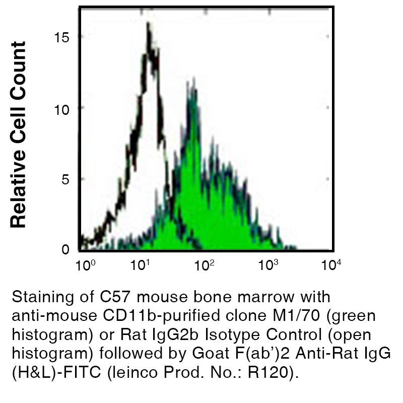
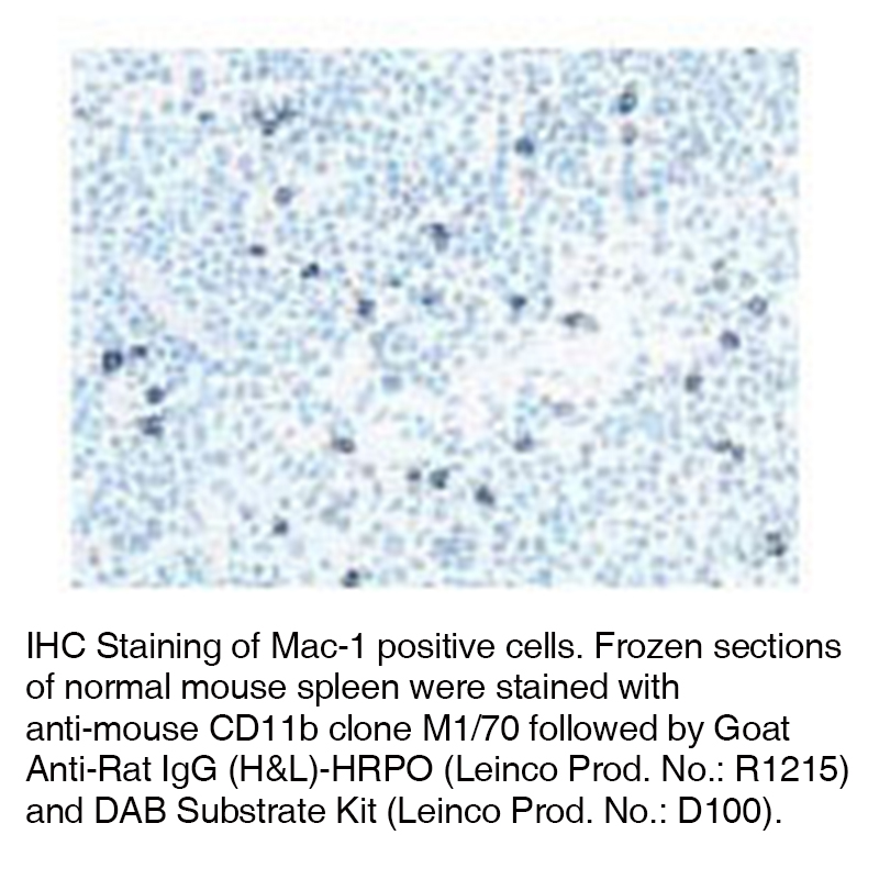
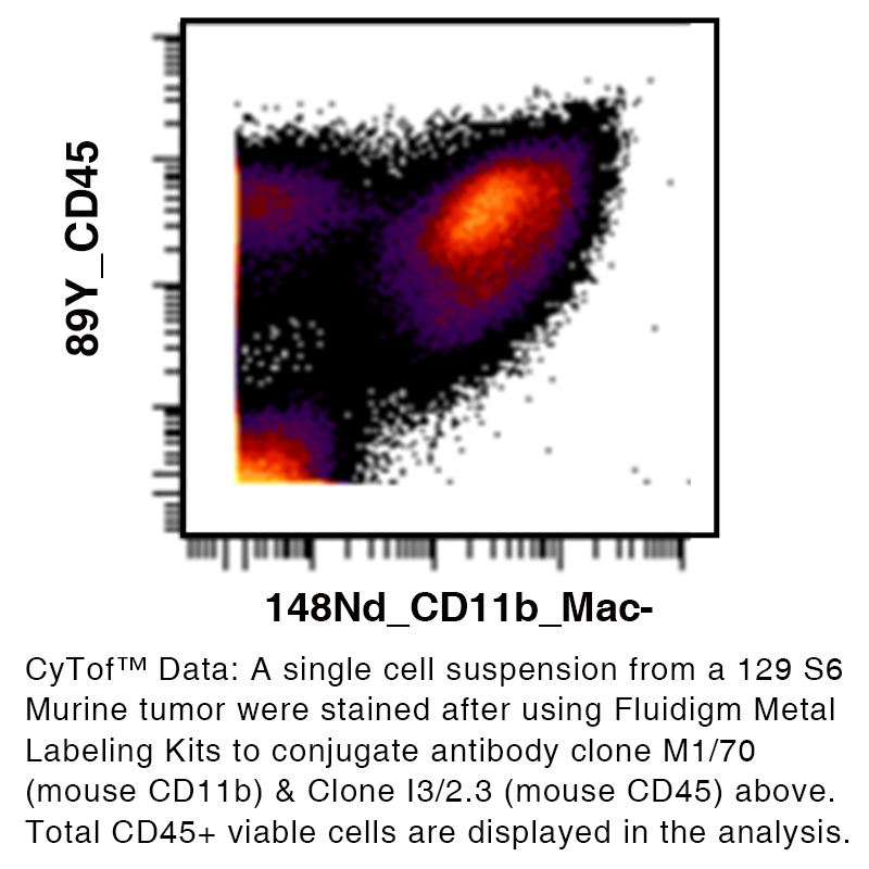
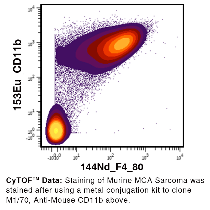
Data
Antibody DetailsProduct DetailsReactive Species Mouse Host Species Rat Recommended Isotype Controls Recommended Isotype Controls Recommended Dilution Buffer Immunogen B10 mouse spleen cells enriched for T cells Product Concentration ≥ 5.0 mg/ml Endotoxin Level <0.5 EU/mg as determined by the LAL method Purity ≥98% monomer by analytical SEC ⋅ >95% by SDS Page Formulation This monoclonal antibody is aseptically packaged and formulated in 0.01 M phosphate buffered saline (150 mM NaCl) PBS pH 7.2 - 7.4 with no carrier protein, potassium, calcium or preservatives added. Due to inherent biochemical properties of antibodies, certain products may be prone to precipitation over time. Precipitation may be removed by aseptic centrifugation and/or filtration. Product Preparation Functional grade preclinical antibodies are manufactured in an animal free facility using in vitro cell culture techniques and are purified by a multi-step process including the use of protein A or G to assure extremely low levels of endotoxins, leachable protein A or aggregates. Pathogen Testing To protect mouse colonies from infection by pathogens and to assure that experimental preclinical data is not affected by such pathogens, all of Leinco’s Purified Functional PLATINUM™ antibodies are tested and guaranteed to be negative for all pathogens in the IDEXX IMPACT I Mouse Profile. Storage and Handling Functional grade preclinical antibodies may be stored sterile as received at 2-8°C for up to one month. For longer term storage, aseptically aliquot in working volumes without diluting and store at ≤ -70°C. Avoid Repeated Freeze Thaw Cycles. Country of Origin USA Shipping Next Day 2-8°C RRIDAB_2829819 Applications and Recommended Usage? Quality Tested by Leinco FC The suggested concentration for this M1/70 antibody for staining cells in flow cytometry is ≤ 0.25 μg per 106 cells in a volume of 100 μl. Titration of the reagent is recommended for optimal performance for each application. Additional Applications Reported In Literature ? CyTOF® PhenoCycler-Fusion (CODEX)® ICC IHC FF Additional Reported Applications For Relevant Conjugates ? B Depletion FC ICC IHC (Frozen) IHC (Paraffin) IP For specific conjugates of this clone, review literature for suggested application details. Each investigator should determine their own optimal working dilution for specific applications. See directions on lot specific datasheets, as information may periodically change. DescriptionDescriptionSpecificity Clone M1/70 recognizes the α subunit (CD11b) of the mouse CD11b/CD18 complex. Background LFA-1α (CD11a) and CD18 are the Integrin alpha-L and beta-2 chains respectively that combine to form LFA-1, a glycoprotein and a member of the Integrin family. Integrin alpha-L/beta-2 is a receptor for ICAM1, ICAM2, ICAM3, ICAM4 and for F11R. LFA-1 participates in the immunological synapses between CD8+ T lymphocytes and antigen-presenting cells. The absence of LFA-1α or ß may induce LAD. The antigen contributes to natural killer cell cytotoxicity, and is involved in various immune phenomena such as leukocyte-endothelial cell interaction, cytotoxic T-cell mediated killing, and antibody dependent killing by granulocytes and monocytes. The CD11b/CD18 antigen is a heterodimeric surface glycoprotein on leukocytes and belongs to the ß2 integrin family. CD11b functions as a receptor for C3bi complement, clotting factor X, fibrinogen and ICAM-1. CD11c forms an α/ß heterodimeric glycoprotein (CD11c/CD18 complex) which belongs to the ß2 integrin family. The complex binds fibrinogen and reportedly serves as a receptor for iC3b and ICAM-1. During inflammatory responses, it mediates cell to cell interaction and is important in both monocyte adhesion and chemotaxis. Antigen Distribution CD11b is expressed on human peripheral blood lymphocytes, NK lymphocytes, monocytes, granulocytes, a subset of T-cells and macrophages. Ligand/Receptor ICAM-1 (CD54), ICAM-2 (CD102), ICAM-4 (CD242), iC3b, fibrinogen Function Adhesion, chemotaxis PubMed NCBI Gene Bank ID UniProt.org Research Area Cell Adhesion . Cell Biology . Costimulatory Molecules . Immunology . Innate Immunity . Neuroscience . Neuroscience Cell Markers Leinco Antibody AdvisorPowered by AI: AI is experimental and still learning how to provide the best assistance. It may occasionally generate incorrect or incomplete responses. Please do not rely solely on its recommendations when making purchasing decisions or designing experiments. Clone M1/70 is widely used in in vivo mouse studies for several applications related to CD11b-expressing cells, such as macrophages, monocytes, neutrophils, and some dendritic cells and NK cells. Common in vivo applications include:
The M1/70 antibody targets CD11b (Integrin alpha M, ITGAM), a common marker for monocytes, macrophages, neutrophils, microglia, and other myeloid cells. In the literature, several other antibodies and proteins are routinely used alongside M1/70, depending on the application (e.g., flow cytometry, immunohistochemistry, lineage depletion, or cell subset identification). Commonly used antibodies or proteins with M1/70 include:
Typical combinations by research context:
Citations in the literature frequently describe panels including several of the above markers, adapted for tissue specificity and purpose. Key point: The specific choice of accompanying antibodies depends on the target cell population, the tissue source, and the experimental question, but CD11b (M1/70) is rarely used in isolation in modern immunological studies. Clone M1/70 is a monoclonal antibody primarily targeting CD11b (integrin alpha-M), widely cited in scientific literature for its role in immunological research. The key findings from M1/70 citations are:
Summary table of key findings:
These findings establish clone M1/70 as an essential tool in immunology for dissecting cell lineage, monitoring inflammatory responses, and blocking integrin-mediated cell functions. Overview of Clone M1/70 (Anti-CD11b) Dosing Regimens in Mouse ModelsClone M1/70 is a widely used rat monoclonal antibody targeting CD11b (integrin alpha M/Mac-1) in mice, primarily employed for myeloid cell identification, functional blocking, and cell subset characterization in immunology, neuroscience, and cancer research. However, detailed, standardized dosing regimens for in vivo use of M1/70 are not consistently published across vendors or in primary literature, unlike other commonly used antibodies (e.g., anti-PD-1, anti-Ly6G). Current Knowledge from Available Sources
Practical Guidance
Summary Table: M1/70 Dosing Information by Application
Key Takeaways
In summary, while M1/70 is a standard tool for myeloid cell identification and functional studies in mice, its in vivo dosing lacks consensus and must be tailored to the specific experimental design and mouse model. References & Citations1. Springer, T.A., et al. (1978) Eur. J. Immunol. 8:539
2. Springer, T.A., et al. (1982) Immunol. Rev. 68:171
3. Ding, A., et al. (1987) J Exp Med. 165:733
4. Vignali, D. A. et al. (1990) J. Immunol. 144:4030
5. Kantor, A. et al. (1992) Proc. Natl. Acad. Sci. U.S.A. 89:3320 Technical ProtocolsCyTOF® FA  ICC IHC FF  PhenoCycler® Certificate of Analysis |
Related Products
Prod No. | Description |
|---|---|
S211 | |
R1364 | |
I-1034 | |
C247 | |
F1175 | |
R1214 | |
S571 |
Formats Available
Prod No. | Description |
|---|---|
C1906 | |
C341 | |
C227 | |
C228 | |
C230 | |
C229 | |
C1628 | |
C1629 | |
C1630 | |
C1632 | |
C515 | |
C377 | |
C677 |
 Products are for research use only. Not for use in diagnostic or therapeutic procedures.