

Anti-Mouse CD32/CD16 [Clone 2.4G2] — Purified in vivo GOLD™ Functional Grade


Anti-Mouse CD32/CD16 [Clone 2.4G2] — Purified in vivo GOLD™ Functional Grade
Product No.: C381
Clone 2.4G2 Target CD32/CD16 Formats AvailableView All Product Type Monoclonal Antibody Alternate Names Fcγ R III/II, Ly-17 Isotype Rat IgG2b Applications B , FA , FC , IHC FF , in vivo , IP , PhenoCycler® , WB |
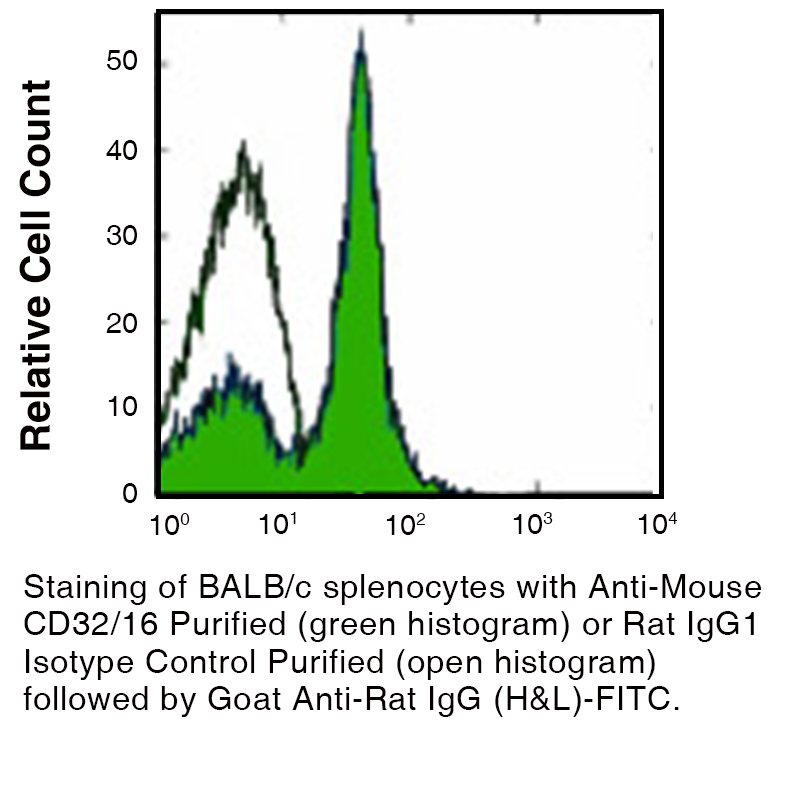
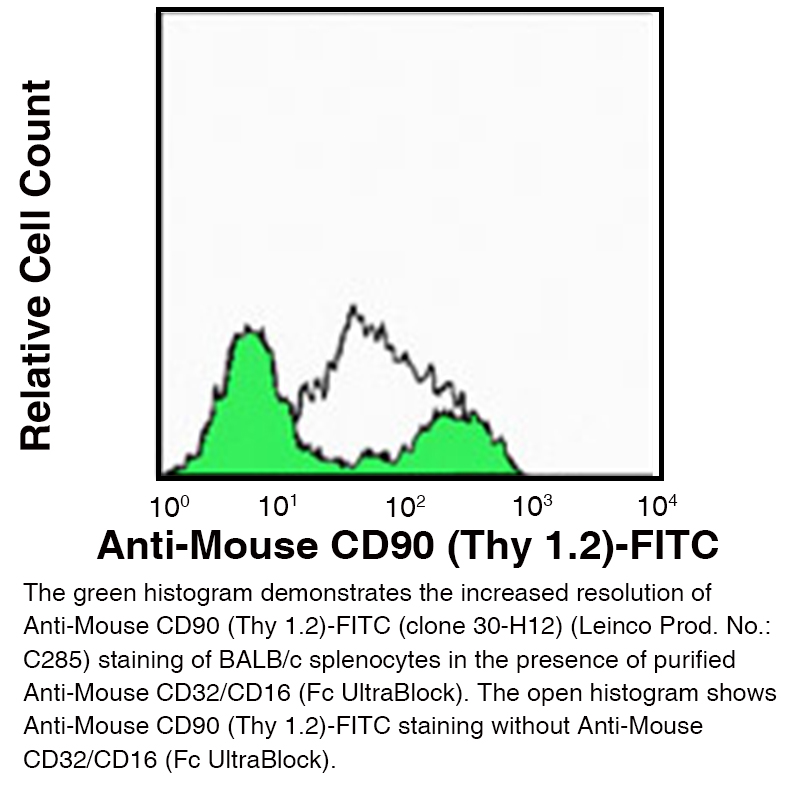
Data
Antibody DetailsProduct DetailsReactive Species Mouse Host Species Rat Recommended Isotype Controls Recommended Isotype Controls Recommended Dilution Buffer Immunogen Sorted pre-B cells Product Concentration ≥ 5.0 mg/ml Endotoxin Level < 1.0 EU/mg as determined by the LAL method Purity ≥95% monomer by analytical SEC ⋅ >95% by SDS Page Formulation This monoclonal antibody is aseptically packaged and formulated in 0.01 M phosphate buffered saline (150 mM NaCl) PBS pH 7.2 - 7.4 with no carrier protein, potassium, calcium or preservatives added. Due to inherent biochemical properties of antibodies, certain products may be prone to precipitation over time. Precipitation may be removed by aseptic centrifugation and/or filtration. Product Preparation Functional grade preclinical antibodies are manufactured in an animal free facility using in vitro cell culture techniques and are purified by a multi-step process including the use of protein A or G to assure extremely low levels of endotoxins, leachable protein A or aggregates. Storage and Handling Functional grade preclinical antibodies may be stored sterile as received at 2-8°C for up to one month. For longer term storage, aseptically aliquot in working volumes without diluting and store at ≤ -70°C. Avoid Repeated Freeze Thaw Cycles. Country of Origin USA Shipping Next Day 2-8°C RRIDAB_2737484 Applications and Recommended Usage? Quality Tested by Leinco FC The suggested concentration for this 2.4G2 antibody for staining cells in flow cytometry is ≤ 1 μg per 106 cells in a volume of 100 μl. Titration of the reagent is recommended for optimal performance for each application. WB The suggested concentration for this 2.4G2 antibody for use in western blotting is 1-10 μg/ml. Additional Applications Reported In Literature ? CODEX® Additional Reported Applications For Relevant Conjugates ? B IP For specific conjugates of this clone, review literature for suggested application details. Each investigator should determine their own optimal working dilution for specific applications. See directions on lot specific datasheets, as information may periodically change. DescriptionDescriptionSpecificity Clone 2.4G2 recognizes the FcγIII and FcγII receptors. Background CD16 is a low affinity Fc gamma receptor that exists in two major forms, CD16a and CD16b, and plays a central role in innate and adaptive immune responses involving antibody‑coated targets. CD16a (FcγRIIIA) is a 50–65 kD transmembrane receptor expressed primarily on natural killer (NK) cells, subsets of T cells, monocytes, and macrophages, where it mediates antibody‑dependent cell‑mediated cytotoxicity (ADCC) and contributes to phagocytosis. CD16b (FcγRIIIB) is a ~48 kD GPI‑anchored receptor expressed mainly on neutrophils; its extracellular domain is over 95% homologous to that of CD16a, enabling efficient binding to IgG immune complexes on pathogen or tumor cell surfaces. Through these interactions, CD16 participates in NK cell activation, triggers downstream signal transduction pathways, and coordinates inflammatory effector functions. CD32 (FcγRII) and CD16 (FcγRIII) together represent key low affinity Fc gamma receptors that recognize antibody–antigen immune complexes and help bridge innate and adaptive immunity. These receptors, typically in the 40–65 kD range depending on isoform and glycosylation state, regulate critical effector processes such as phagocytosis, respiratory burst, cytokine production, and ADCC in response to IgG‑opsonized targets. Because CD16 and CD32 can bind the Fc portion of many commonly used IgG antibodies, blocking CD16/CD32 (FcγRIII/II) is widely used in flow cytometry, immunofluorescence, and other immunoassays to reduce nonspecific Fc‑mediated binding, improve staining specificity, and enhance the reliability of immune cell phenotyping and functional studies. Antigen Distribution These receptors are present on B cells, monocyte/macrophages, NK cells, neutrophils, mast cells and dendritic cells. Ligand/Receptor IgG Function Low affinity receptors for IgG PubMed UniProt.org Research Area Immunology . Innate Immunity Leinco Antibody AdvisorPowered by AI: AI is experimental and still learning how to provide the best assistance. It may occasionally generate incorrect or incomplete responses. Please do not rely solely on its recommendations when making purchasing decisions or designing experiments. Clone 2.4G2 is primarily used in vivo in mice as an antibody to block Fc gamma receptors CD16 (FcγRIII) and CD32 (FcγRII), thereby preventing non-specific binding of experimental antibodies and modulating immune responses. Common in vivo applications include:
Dosage and administration:
Limitations:
Summary table:
2.4G2 is considered a standard tool for Fc receptor research in mouse immunology. Commonly used antibodies and proteins paired with 2.4G2 (anti-mouse CD16/CD32) in the literature include cell surface marker antibodies for multiparameter flow cytometry—specifically B220, CD3, CD19, CD11b, Gr1, and NK1.1. These combinations enable precise immune cell phenotyping by blocking nonspecific Fc receptor-mediated antibody binding. Most frequently co-used antibodies/proteins:
Other frequent combinations and controls:
Technical practices:
Detection and analysis applications:
Researchers may also use other Fc receptor-blocking antibodies, such as clone 93, though it is distinct from 2.4G2 and not interchangeable in all experiments. Summary Table:
In summary, 2.4G2 is typically used in combination with a broad set of lineage-defining antibodies and appropriate controls in multiparametric mouse immunophenotyping and functional immune assays, with attention to the technical details needed to ensure specific and accurate results. Clone 2.4G2 is a monoclonal antibody extensively cited in scientific literature for its role as a blocker of mouse Fc gamma receptors II and III (FcγRII/CD32 and FcγRIII/CD16). Its key findings and uses include:
In summary, clone 2.4G2’s chief scientific value is its reproducible, efficient blockade of FcγRII and FcγRIII, minimizing non-specific antibody binding and enabling high-quality immunological research and functional studies in mice. Dosing regimens of clone 2.4G2 (anti-mouse CD32/CD16) are largely standardized across different mouse models, most commonly employing a single 500 µg intraperitoneal (i.p.) injection 24 hours prior to experimental intervention. Significant variations in dose, schedule, or administration route are rare and not routinely reported in the literature. Key details:
Summary dosing table for commonly used models:
Additional notes:
In summary, while minor adjustments may occur based on experimental nuance, clone 2.4G2 dosing is highly consistent across mouse models, with 500 µg i.p. 24h before intervention as the gold standard for acute receptor blockade. References & Citations1.) Titas, J. A. et al. (1982) J. Immunol. 133:556 2.) Rodewald, H. et al. (1992) Cell 69:139 3.) Skyberg, J. A. et al. (2020) Infection and Immunity. 88: 5 4.) Forte et al. (2020) Cell Reports. 30:3149–3163 Journal Link 5.) Forte, E. et al. (2020) Cell Reports 30(9):3149-3163.e6 Journal Link Technical ProtocolsB FA  IHC FF   PhenoCycler®  Certificate of Analysis |
Related Products
Prod No. | Description |
|---|---|
S211 | |
R1364 | |
I-1034 | |
C247 | |
F1175 | |
R1214 | |
S571 |
Formats Available
Prod No. | Description |
|---|---|
C2078 | |
C2080 | |
C2084 | |
C524 | |
C381 | |
C247 | |
C250 | |
C2082 | |
C348 | |
C681 | |
C248 | |
C249 |
 Products are for research use only. Not for use in diagnostic or therapeutic procedures.