Anti-Mouse CD86 [Clone GL1] — Purified in vivo PLATINUM™ Functional Grade
Anti-Mouse CD86 [Clone GL1] — Purified in vivo PLATINUM™ Functional Grade
Product No.: C6158
Clone GL1 Target B7-2 Formats AvailableView All Product Type Monoclonal Antibody Alternate Names B7-2, B70, Ly-58, CD-86 Isotype Rat IgG2a κ Applications B , ELISA , FC , IHC FF , in vivo , IP , WB |
Data
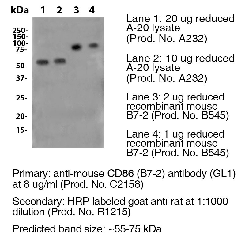
Antibody DetailsProduct DetailsReactive Species Mouse Host Species Rat Recommended Isotype Controls Recommended Isotype Controls Recommended Dilution Buffer Immunogen LPS-activated CBA/Ca mouse splenic B cells Product Concentration ≥ 5.0 mg/ml Endotoxin Level <0.5 EU/mg as determined by the LAL method Purity ≥98% monomer by analytical SEC ⋅ >95% by SDS Page Formulation This monoclonal antibody is aseptically packaged and formulated in 0.01 M phosphate buffered saline (150 mM NaCl) PBS pH 7.2 - 7.4 with no carrier protein, potassium, calcium or preservatives added. Due to inherent biochemical properties of antibodies, certain products may be prone to precipitation over time. Precipitation may be removed by aseptic centrifugation and/or filtration. Product Preparation Functional grade preclinical antibodies are manufactured in an animal free facility using in vitro cell culture techniques and are purified by a multi-step process including the use of protein A or G to assure extremely low levels of endotoxins, leachable protein A or aggregates. Pathogen Testing To protect mouse colonies from infection by pathogens and to assure that experimental preclinical data is not affected by such pathogens, all of Leinco’s Purified Functional PLATINUM™ antibodies are tested and guaranteed to be negative for all pathogens in the IDEXX IMPACT I Mouse Profile. Storage and Handling Functional grade preclinical antibodies may be stored sterile as received at 2-8°C for up to one month. For longer term storage, aseptically aliquot in working volumes without diluting and store at ≤ -70°C. Avoid Repeated Freeze Thaw Cycles. Country of Origin USA Shipping Next Day 2-8°C RRIDAB_2829780 Applications and Recommended Usage? Quality Tested by Leinco ELISAThis antibody is useful as the capture antibody in a sandwich ELISA. The suggested coating concentration is 5 µg/ml (100 µl/well) µg/ml. WB This antibody can be used to detect Human, Mouse and Rat TIM-1 by Western blot analysis at a concentration of 1.0-2.0 µg/ml when used in conjunction with compatible secondary reagents, such as PN:R951, under either reducing or non-reducing conditions. The positive control for Western blotting is PN:U124. Additional Applications Reported In Literature ? B Each investigator should determine their own optimal working dilution for specific applications. See directions on lot specific datasheets, as information may periodically change. DescriptionDescriptionSpecificity Clone GL-1 recognizes an epitope on mouse CD86. Background CD86 is an 80kD Ig superfamily member that is involved in immunoglobulin class-switching and activation of NK cell-mediated cytotoxicity. CD80 is closely related to, and works in tandem with CD86 to prime T- cells. CD86 is expressed earlier in the immune response than CD80. The ligation of CD28 on T cells with CD80 and CD86 on APCs co-stimulates T cells resulting in enhanced cell activation, proliferation, and cytokine production. CD86 can also bind to CTLA-4 to deliver an inhibitory signal to T cells. Antigen Distribution CD86 is expressed on activated B and T cells, macrophages, dendritic cells, and astrocytes. Ligand/Receptor CD28, CD152 (CTLA-4) Function T cell costimulation, Ig class-switching, NK cell cytotoxicity PubMed NCBI Gene Bank ID UniProt.org Research Area Cell Biology . Costimulatory Molecules . Immunology . Neuroscience . Neuroscience Cell Markers Leinco Antibody AdvisorPowered by AI: AI is experimental and still learning how to provide the best assistance. It may occasionally generate incorrect or incomplete responses. Please do not rely solely on its recommendations when making purchasing decisions or designing experiments. Clone GL1 is most commonly used in vivo in mice to block CD86 (B7-2)-dependent costimulatory signaling, thereby inhibiting T cell-mediated immune responses and enabling mechanistic studies of immune regulation. Key in vivo applications include:
Additional notes:
In summary, the principal in vivo use of clone GL1 in mice is as a CD86-blocking antibody to dissect and modulate T cell-dependent immune processes, enabling both basic immunological research and preclinical modeling of disease and immunotherapy. Antibodies and Proteins Commonly Used with GL1 in the LiteratureGL1 is a research tool, specifically the CD86 (B7-2) Monoclonal Antibody clone GL1, used to recognize and manipulate the CD86 protein, which is a co-stimulatory molecule involved in T cell activation. There is limited published information directly pairing GL1 with other specific antibodies or proteins in functional assays beyond its standard use to detect CD86. However, we can outline the typical experimental context and related reagents, as well as clarify the distinction with unrelated abbreviations (such as GLUT1 or GLP-1). Context of GL1 (CD86) Use
Example Experimental Combinations
Distinction from GLUT1, GLP-1, and Other AbbreviationsSome literature refers to GLUT1 (glucose transporter 1) antibodies or GLP-1 (glucagon-like peptide-1) receptor agonists, which are unrelated to CD86 or GL1. These are not used in combination with GL1 (CD86) but rather in metabolic or diabetes research. For example, GLUT1 antibodies target the glucose transporter for structural or functional studies in cell membranes, while GLP-1 agonists are peptide-based drugs for diabetes and obesity. Summary
If you have a more specific experimental context or are referring to a different "GL1" system, please clarify for a tailored response. The key findings from clone GL1 citations in scientific literature vary significantly depending on the research context, as this designation appears in both immunological and metabolic research domains. GLP-1 Analogue Production and Therapeutic DevelopmentIn the context of GLP-1 (glucagon-like peptide-1) research, clone GL1 citations focus on advances in recombinant production, bioactivity optimization, and molecular engineering for therapeutic applications in diabetes and obesity. A recent breakthrough involved engineering a high-throughput clone for industrial-scale production of long-acting GLP-1 analogues with retained bio-efficacy. This work demonstrated that incorporating n-terminal hydrophobic amino acids, thioredoxin-modified tags, and enterokinase cleavage sites resulted in a 1.5-fold increase in mRNA gene expression, achieving final product yields of 170-190 mg per liter of fermentation culture. The therapeutic development of GLP-1-based drugs represents a major milestone in medicine, solving a century-old mystery about the gut's role in glucose metabolism. The discovery that GLP-1(7-37) is produced in the intestine and functions as an incretin hormone laid the foundation for revolutionary treatments for metabolic disease. Recent clinical research has shown that GLP-1 receptor agonists may reduce the risk of specific obesity-associated cancers compared with insulins in patients with type 2 diabetes. Immunological ApplicationsIn immunology research, clone GL1 refers to an anti-mouse CD86 antibody used primarily in in vivo mouse studies to block CD86-dependent costimulatory signaling. CD86 plays a crucial role in T cell activation by binding to CD28 on T cells, resulting in enhanced cell activation, proliferation, and cytokine production. This clone recognizes an epitope on mouse CD86, an 80kD immunoglobulin superfamily member involved in immunoglobulin class-switching and activation of NK cell-mediated cytotoxicity. The antibody is used to inhibit T cell-mediated immune responses by blocking the CD86-CD28 costimulatory pathway. Dosing regimens for clone GL1 (an anti-mouse CD86 [B7-2] monoclonal antibody) are not standardized across all mouse models; they are typically adjusted depending on experimental objectives, route of administration (intravenous, intraperitoneal, or in vivo blocking), and the immune context of the model. Protocols published by suppliers and in research literature offer some guidance:
How dosing may vary by mouse model:
For example, the Leinco and BioLegend datasheets suggest:
Summary Table—Dosing Regimens for GL1 Clone (Anti-mouse CD86):
Explicit adjustments should be guided by pilot data and validated in the specific model. Always refer to product datasheets for starting recommendations and consult primary literature for models matching your application. Currently, the literature and datasheets do not report comprehensive pharmacokinetic or strain-dependent dosing studies for GL1 specifically; thus, empirical optimization is standard practice. References & Citations1. Hathcock, K.S. et al.. (1993) Science 262(5135:905-7 Technical ProtocolsCertificate of Analysis |
Related Products
Prod No. | Description |
|---|---|
S211 | |
R1364 | |
I-1177 | |
C247 | |
F1175 | |
R1214 | |
S571 |
Formats Available
Prod No. | Description |
|---|---|
C2168 | |
C2163 | |
C2164 | |
C2165 | |
C2166 | |
C2167 | |
C2159 | |
C2160 | |
C2161 | |
C2162 | |
C2158 | |
C2050 | |
C6158 | |
B585 |
 Products are for research use only. Not for use in diagnostic or therapeutic procedures.


