

Anti-Mouse CD172a [P84] – Purified in vivo PLATINUM™ Functional Grade


Anti-Mouse CD172a [P84] – Purified in vivo PLATINUM™ Functional Grade
Product No.: P680
Clone P84 Target CD172a Formats AvailableView All Product Type Monoclonal Antibody Alternate Names SHPS-1, BIT, P84, PTPNS1, CD172 antigen-like family member A Isotype Rat IgG1 κ Applications B , CyTOF® , FC , IHC FF , in vivo , IP |
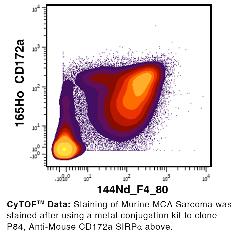
Data
Antibody DetailsProduct DetailsReactive Species Mouse Host Species Rat Recommended Isotype Controls Recommended Isotype Controls Recommended Dilution Buffer Immunogen Mouse brain membrane protein Product Concentration ≥ 5.0 mg/ml Endotoxin Level <0.5 EU/mg as determined by the LAL method Purity ≥98% monomer by analytical SEC ⋅ >95% by SDS Page Formulation This monoclonal antibody is aseptically packaged and formulated in 0.01 M phosphate buffered saline (150 mM NaCl) PBS pH 7.2 - 7.4 with no carrier protein, potassium, calcium or preservatives added. Due to inherent biochemical properties of antibodies, certain products may be prone to precipitation over time. Precipitation may be removed by aseptic centrifugation and/or filtration. Product Preparation Functional grade preclinical antibodies are manufactured in an animal free facility using in vitro cell culture techniques and are purified by a multi-step process including the use of protein A or G to assure extremely low levels of endotoxins, leachable protein A or aggregates. Pathogen Testing To protect mouse colonies from infection by pathogens and to assure that experimental preclinical data is not affected by such pathogens, all of Leinco’s Purified Functional PLATINUM™ antibodies are tested and guaranteed to be negative for all pathogens in the IDEXX IMPACT I Mouse Profile. Storage and Handling Functional grade preclinical antibodies may be stored sterile as received at 2-8°C for up to one month. For longer term storage, aseptically aliquot in working volumes without diluting and store at ≤ -70°C. Avoid Repeated Freeze Thaw Cycles. Country of Origin USA Shipping Next Day 2-8°C Working Concentration 1.0 mg/ml RRIDAB_2831659 Applications and Recommended Usage? Quality Tested by Leinco FC The suggested concentration for this P84 antibody for staining cells in flow cytometry is ≤ 1.0 μg per 106 cells in a volume of 100 μl. Titration of the reagent is recommended for optimal performance for each application. CyTOF® Additional Applications Reported In Literature ? IHC FF IP B Each investigator should determine their own optimal working dilution for specific applications. See directions on lot specific datasheets, as information may periodically change. DescriptionDescriptionSpecificity Clone P84 recognizes an epitope on mouse CD172a.
Background CD172a antibody, clone P84, recognizes CD172a, also known as single regulatory protein α (SIRPα) (signal regulatory protein alpha) or Src homology 2 domain-containing phosphatase substrate-1 (SHP-1), a type I transmembrane glycoprotein with three Ig-like extracellular domains and two cytoplasmic immunoreceptor tyrosine-based inhibition motifs (ITIMs)1. SIRPα is expressed predominantly in myeloid cells2 - including monocytes, macrophages, and dendritic cells (DCs) - and neuronal cells3. The extracellular ligand for SIRPα, CD47 (or integrin-associated protein [IAP])4, is expressed in most cell types5. In macrophages, ligation of SIRPα by CD47 inhibits macrophage phagocytosis of self cells6,7. SIRPα also negatively regulates DC-mediated T cell activation and DC maturation8-10. CD47 is also upregulated on tumor cells, inhibiting the phagocytosis of tumor cells by macrophages11. Therapeutics targeting the CD47-SIRPα interaction, including antibodies and fusion proteins, are currently under preclinical and clinical study for various malignancies as a monotherapy or in combination with other therapeutics12. Antigen Distribution CD172a is expressed on monocytes, macrophages, dendritic cells, and neuronal cells.
Ligand/Receptor CD47, SP-A, SP-D Function Negative regulation of several biological processes NCBI Gene Bank ID UniProt.org Research Area Cell Biology . Immunology Leinco Antibody AdvisorPowered by AI: AI is experimental and still learning how to provide the best assistance. It may occasionally generate incorrect or incomplete responses. Please do not rely solely on its recommendations when making purchasing decisions or designing experiments. Common In Vivo Applications of Clone P84 in MiceClone P84 is a widely used rat monoclonal antibody targeting mouse CD172a (SIRPα), a receptor expressed predominantly on myeloid cells including monocytes, macrophages, dendritic cells, and some neurons. Its in vivo utility is supported by several key applications:
Technical Considerations
Summary Table
Clone P84 is thus a versatile tool for in vivo studies focused on the SIRPα–CD47 axis, particularly in cancer immunotherapy, immune modulation, and basic biology research in mice. Commonly used antibodies or proteins alongside P84 (often referring to the anti-mouse CD172a [P84] antibody or the nuclear matrix protein p84 antibody) include several secondary reagents, cellular markers, and multiplexed targets for broader biological context or multiplex analysis. Key examples from the literature include:
Overview Table:
The exact set of co-used antibodies or proteins will vary based on experiment type (IHC, IF, IP, flow cytometry) and biological system under study. Clone P84 is a rat monoclonal antibody that specifically recognizes CD172a (SIRPα - Signal Regulatory Protein alpha) in mice, and research using this antibody has revealed several important findings about SIRPα function and its potential therapeutic applications. Molecular Characterization of P84/SIRPαThe P84 molecule was identified as being identical to SHPS-1, a member of the immunoglobulin superfamily. The full-length cDNA encodes a 509-amino-acid peptide with a calculated molecular weight of 56 kDa, though it appears larger on SDS-PAGE due to extensive glycosylation through 17 potential N-glycosylation sites. When treated with N-glycosidase F, the molecular weight shifts from 86 kDa to 64 kDa and from 77 kDa to 42-55 kDa, confirming the significant contribution of carbohydrate modifications. The protein exists in two forms generated by alternative splicing, with the smaller form lacking a 218-amino-acid segment. The molecule contains a transmembrane domain and a cytoplasmic segment with multiple potential tyrosine phosphorylation sites at positions 436, 460, 477, and 501, which are crucial for its signaling function. These phosphorylation sites enable the recruitment and activation of tyrosine phosphatases SHP-1 and SHP-2 through immunoreceptor tyrosine-based inhibitory motifs (ITIMs). Cancer Immunotherapy ApplicationsResearch using clone P84 has demonstrated significant implications for cancer therapy. Studies comparing P84 with another anti-SIRPα antibody (MY-1) revealed important functional differences in blocking the CD47-SIRPα interaction. While MY-1 blocks CD47 binding and showed marked attenuation of tumor formation, P84 has little effect on the CD47-SIRPα interaction but still demonstrated a smaller yet significant inhibitory effect on tumor growth in RENCA renal cell carcinoma models. When mice were treated with P84 after tumor establishment, the antibody showed reduced efficacy compared to early treatment, with delayed treatment having no significant effect on either tumor growth or survival. In metastatic models using B16BL6 melanoma cells, P84 was less effective than MY-1 in reducing lung metastatic nodules. These findings suggest that antibodies blocking the CD47-SIRPα interaction are more effective than those that don't, though P84 retains some tumor-suppressive activity through alternative mechanisms. Cellular Expression and FunctionClone P84 has been extensively used to characterize SIRPα expression patterns on myeloid cells, including granulocytes, dendritic cells, macrophages, mast cells, and hematopoietic stem cells. The antibody has proven particularly useful in flow cytometric analysis of mouse bone marrow cells, where it helps identify CD11b+ myeloid populations expressing CD172a. Studies have shown that SIRPα plays regulatory roles in several physiological processes, including inhibition of host cell phagocytosis by macrophages and bi-directional activation of T cells and dendritic cells. The protein is involved in negative regulation of receptor tyrosine kinase-coupled signaling pathways and responds to various stimuli including serum, growth factors, insulin, and cell adhesion signals. Technical ApplicationsClone P84 has been reported to have neutralizing activity and is widely used in flow cytometry applications. The antibody can be used at concentrations of 0.5 µg or less per test, with optimal detection using blue, green, or yellow-green lasers with excitation at 488-561 nm. It demonstrates specificity for SIRPα without cross-reactivity to SIRPβ family members, unlike some other anti-SIRPα antibodies. The antibody is available in multiple formats including purified, PE-conjugated, and Ultra-LEAF preparations for in vivo functional studies. Clone P84 has become a standard research tool for investigating SIRPα biology and evaluating therapeutic strategies targeting the CD47-SIRPα axis in cancer and other diseases. Dosing Regimens of Clone P84 (Anti-Mouse CD172a/SIRPα) in Mouse ModelsClone P84 is a monoclonal antibody targeting mouse CD172a, also known as Signal Regulatory Protein Alpha (SIRPα), and is widely used for in vivo experiments, flow cytometry, and functional studies in mice. The dosing regimens in mouse models vary depending on the experimental design, target tissue, and therapeutic or mechanistic goals. Key Examples of Dosing RegimensThe available literature provides several specific examples of how P84 is administered in different models, with dosing that is tailored to the experimental context: Neuroblastoma Tumor Model (NXS2)
Staining for Flow Cytometry
Bulk In Vivo Antibody Supply
Additional Notes
Summary Table
ConclusionDosing regimens for clone P84 vary considerably across mouse models, with published in vivo regimens typically around 20 μg per mouse per injection for therapeutic studies, while flow cytometry uses much smaller amounts per cell. The dosing should be tailored to the specific experimental endpoint, and researchers are encouraged to consult both published literature and antibody provider protocols for optimal use. For new or specialized models, dose-finding studies may still be necessary. References & Citations1. Fujioka, Y., et al. (1996) Mol. Cell. Biol. 16:6887 2. Adams, S., et al. (1998) J. Immunol. 161:1853 3. Chuang W, et al. (1990) Dev Biol. 137:219–232 4. Seiffert, M., et al. (1999) Blood 94:3633 5. Oldenborg P. A. (2013) ISRN Hematol. 2013:614619 6. Oldenborg, P. A., et al. (2000) Science 288:2051 7. Oldenborg, P. A., et al. (2001) J. Exp. Med. 193:855 8. Brooke, G. P., et al. (1998) Eur. J. Immunol. 28:1 9. Seiffert, M., et al. (2001) Blood 97:2741 10. Latour, S., et al. (2001) J. Immunol. 167:2547 11. Jaiswal S, et al. (2009) Cell. 138(2):271-85 12. Jalil AR, et al. (2020) Antib Ther. 3(2):80-94 Technical ProtocolsB CyTOF®  IHC FF   Certificate of Analysis |
Related Products
Prod No. | Description |
|---|---|
S211 | |
R1364 | |
I-1195 | |
C247 | |
F1175 | |
R1214 | |
S571 |
Formats Available
Prod No. | Description |
|---|---|
P386 | |
P387 | |
P380 | |
P390 | |
P391 | |
P392 | |
P385 | |
P389 | |
P388 | |
P680 | |
S200 |
 Products are for research use only. Not for use in diagnostic or therapeutic procedures.