

Anti-Mouse/Human CD44 [Clone IM7] — Purified in vivo GOLD™ Functional Grade


Anti-Mouse/Human CD44 [Clone IM7] — Purified in vivo GOLD™ Functional Grade
Product No.: C382
Clone IM7 Target CD44 Formats AvailableView All Product Type Monoclonal Antibody Alternate Names Hermes, Pgp-1, H-CAM, HUTCH-1, ECMR III, gp85, Ly-24 Isotype Rat IgG2b κ Applications CyTOF® , ELISA Det , FC , ICC , IHC FF , IHC FFPE , in vivo , IP , PhenoCycler® , WB |
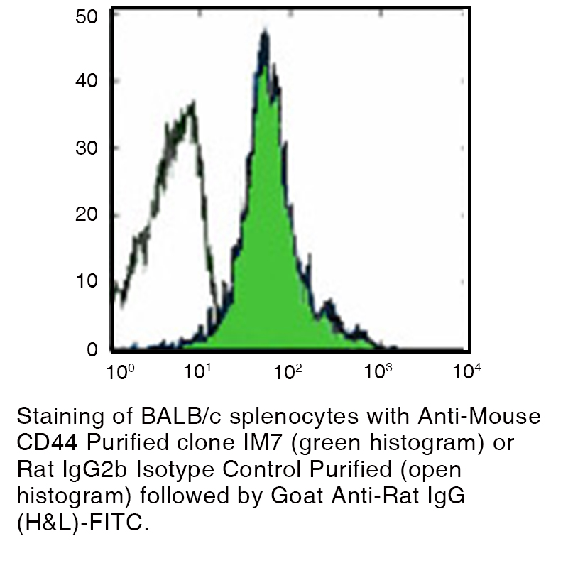
Data
Antibody DetailsProduct DetailsReactive Species Human ⋅ Mouse Host Species Rat Recommended Isotype Controls Recommended Isotype Controls Recommended Dilution Buffer Immunogen Dexamethasone-induced myeloid leukemia M1 cells Product Concentration ≥ 5.0 mg/ml Endotoxin Level < 1.0 EU/mg as determined by the LAL method Purity ≥95% monomer by analytical SEC ⋅ >95% by SDS Page Formulation This monoclonal antibody is aseptically packaged and formulated in 0.01 M phosphate buffered saline (150 mM NaCl) PBS pH 7.2 - 7.4 with no carrier protein, potassium, calcium or preservatives added. Due to inherent biochemical properties of antibodies, certain products may be prone to precipitation over time. Precipitation may be removed by aseptic centrifugation and/or filtration. Product Preparation Functional grade preclinical antibodies are manufactured in an animal free facility using in vitro cell culture techniques and are purified by a multi-step process including the use of protein A or G to assure extremely low levels of endotoxins, leachable protein A or aggregates. Storage and Handling Functional grade preclinical antibodies may be stored sterile as received at 2-8°C for up to one month. For longer term storage, aseptically aliquot in working volumes without diluting and store at ≤ -70°C. Avoid Repeated Freeze Thaw Cycles. Country of Origin USA Shipping Next Day 2-8°C RRIDAB_2737485 Applications and Recommended Usage? Quality Tested by Leinco FC The suggested concentration for this IM7 antibody for staining cells in flow cytometry is ≤ 0.25 μg per 106 cells in a volume of 100 μl or 100μl of whole blood. Titration of the reagent is recommended for optimal performance for each application. WB The suggested concentration for this P84 antibody for use in western blotting is 1-10 μg/ml. Additional Applications Reported In Literature ? CyTOF® ELISA Det ICC IP IHC FF IHC FFPE Additional Reported Applications For Relevant Conjugates ? CODEX® Each investigator should determine their own optimal working dilution for specific applications. See directions on lot specific datasheets, as information may periodically change. DescriptionDescriptionSpecificity Clone IM7 recognizes an epitope common to alloantigens and all isoforms of CD44 that is located between amino acids 145 and 186. Background CD44 is an 80-95 kD glycoprotein that plays a role in various cellular functions including lymphocyte activation, recirculation and homing, hematopoiesis, and tumor metastasis. CD44 interacts with osteopontin, collagens, and matrix metalloproteinases (MMPs) and is a receptor for hyaluronic acid. Transcripts for this gene go through intricate alternative splicing that result in a variety of functionally distinct isoforms, including those which may be related to tumor metastasis. These splice variants of CD44 function as receptors under hemodynamic flow conditions that are significant to the development of cancer metastasis. Hence, it is thought that anti-CD44 tumor-specific mAbs may have therapeutic potential. This therapeutic potential of anti-CD44 mAbs is evident in some animal experiments demonstrating a reduction in malignant activities of various neoplasms when CD44 was targeted by a combination of mAbs, antisense oligonucleotides, and CD44-soluble proteins. It has been reported that high levels of CD44 on leukemic cells fuel leukemia production. Notably, various cancer studies show conflicting results pertaining to level of CD44 expression and its correlation with disease prognosis. Before anti-CD44 therapy can be applied to human cancers, it is essential to resolve this inconsistency. Antigen Distribution CD44 is expressed on all leukocytes, endothelial cells, hepatocytes, and mesenchymal cells in addition to B-cells, monocytes, macrophages and certain subsets of thymocytes and peripheral T-cells. Mice with the Ly-24.1 allotype have high densities of CD44+ T-cells. Ligand/Receptor Hyaluronan, MIP-1β, fibronectin, collagen Function Leukocyte attachment and rolling on endothelial cells, stromal cells and ECM PubMed NCBI Gene Bank ID UniProt.org Research Area Cell Adhesion . Cell Biology . Immunology . Stem Cell Leinco Antibody AdvisorPowered by AI: AI is experimental and still learning how to provide the best assistance. It may occasionally generate incorrect or incomplete responses. Please do not rely solely on its recommendations when making purchasing decisions or designing experiments. Common In Vivo Applications of Clone IM7 in MiceAnti-CD44 monoclonal antibody clone IM7 is widely used in vivo to study the function of the CD44 adhesion molecule, which plays pivotal roles in cell migration, lymphocyte activation, immune response modulation, and tumor biology. IM7 binds to an epitope conserved across all known isoforms of mouse and human CD44, making it a versatile tool in immunology and inflammation research. Key In Vivo Uses
Additional Biological Effects Observed
Typical Experimental ContextIM7 is generally administered intravenously or intraperitoneally, and its effects are monitored in various disease models, including arthritis, DTH, and acute shock. The antibody’s ability to recognize all CD44 isoforms and its documented in vivo efficacy make it a mainstay in immunology and inflammation research in mice. Summary Table
In summary, clone IM7 is most commonly used in vivo in mice to modulate immune and inflammatory responses by blocking CD44, with established roles in models of arthritis and DTH, and is occasionally used to study acute systemic reactions and tumor biology. Researchers should be aware of the potential to induce systemic shock in some contexts. Common Antibodies and Proteins Used with IM7 in the LiteratureIM7 refers primarily to two distinct entities in the literature:
Below, the common co-used antibodies/proteins are summarized for each context. IM7 (Anti-CD44 Monoclonal Antibody)The IM7 monoclonal antibody is specific for CD44, a cell adhesion molecule expressed on hematopoietic and non-hematopoietic cells. In experimental settings, IM7 is often used alongside other antibodies targeting cell surface markers, adhesion molecules, or functional reagents, depending on the research focus:
Key Application Contexts: Im7 (Colicin E7 Immunity Protein)Im7 is a bacterial protein that binds tightly to the DNase domain of colicin E7, forming one of the tightest known protein–protein interactions. In protein engineering, Im7 is used as a scaffold for creating novel binding proteins. Common partners include:
Key Application Contexts: Summary Table
Key Points
Clone IM7 is a monoclonal antibody widely cited in scientific literature for its role in recognizing CD44, a cell surface glycoprotein important in cell adhesion, migration, and signaling. The key findings from studies citing clone IM7 include:
In summary, clone IM7 is a gold-standard reagent for CD44 detection, functional analysis, and therapeutic model studies related to immune modulation, cancer, and inflammation, with broad methodological applications and major contributions to the understanding of CD44 biology. Dosing regimens for clone IM7 (anti-CD44) in mouse models generally range from 1–10 mg/kg, most commonly as single or repeat intraperitoneal injections, but the precise regimen varies based on the experimental goal, mouse strain, and disease model. The literature and product protocols highlight variability, with most applications specifying dosing empirically or referencing previous studies rather than a universally fixed protocol. Key details include:
Reference Example Regimen:A commonly cited regimen is 200–250 μg of IM7 per mouse (i.p.), administered once or at intervals (every 3–7 days) for chronic experiments. This is consistent across suppliers (Bio X Cell, Leinco) and published experimental protocols. Considerations Influencing Regimen Choice:
Flow Cytometry/Profiling Use:For flow cytometry and ex vivo tissue profiling (not in vivo blockade/depletion), clone IM7 is typically used at a much lower concentration, often a 1:100 dilution with 1 × 10⁵–1 × 10⁷ cells/mL in the staining buffer. Summary Table: Clone IM7 Dosing Regimens in Mouse Models
For best results, adaptation to experimental requirements and mouse strain is advised, and supplier protocols or prior peer-reviewed studies should be consulted for the specific disease or immune system model in use. References & Citations1. Trowbridge, I. et al. (1982) Immunogenetics 15:299
2. Lesley, J. et al. (1988) Cell Immunol. 112:40 Technical ProtocolsCyTOF® ELISA Det  ICC IHC FF IHC FFPE   PhenoCycler®  Certificate of Analysis |
Related Products
Prod No. | Description |
|---|---|
S211 | |
R1364 | |
I-1034 | |
C247 | |
F1175 | |
R1214 | |
S571 |
Formats Available
Prod No. | Description |
|---|---|
C1894 | |
C349 | |
C251 | |
C252 | |
C253 | |
C254 | |
C1886 | |
C1887 | |
C1888 | |
C1892 | |
C501 | |
C382 | |
C1890 | |
C2368 |
 Products are for research use only. Not for use in diagnostic or therapeutic procedures.